2723
Ultra-fast EPI sampling of pulsatile flow waveforms in cerebral arteries via retrospective binning of k-space lines1CUBRIC, School of Physics and Astronomy, Cardiff University, Cardiff, United Kingdom, 2Siemens Healthcare GmbH, Erlangen, Germany, 3Siemens Healthcare Ltd, Camberly, United Kingdom, 4CUBRIC, School of Engineering, Cardiff, United Kingdom
Synopsis
Flow related signal enhancement in ultra-fast EPI allows imaging of cardiac pulsatile blood flow profiles in cerebral arteries. We present a novel method that uses retrospective binning of k-space lines to make cardiac phase ‘composite’ k-space planes, from which pulsatile waveforms can be reconstructed with extremely high temporal resolution (~2ms). We demonstrate the proof-of-principle for obtaining pulse wave velocity measures in cerebral arteries, paving the way for mapping quantitative arterial stiffness measures across the brain.
Purpose
Arterial stiffness (AS) is a vascular health index with substantial clinical relevance1,2. Although carotid-femoral pulse wave velocity (PWV) is the gold standard measurement in the body, a more localised measure in the brain is desirable to examine the influence of AS on neurological deterioration. Fast EPI can be made sensitive to cerebral flood flow velocity (CBFV)3,4, allowing cardiac pulsatile flow waveforms to be measured, promising the development of a cerebral AS measure. This study has three purposes; 1) to demonstrate that fast EPI acquisitions yield monotonic sensitivity to flow velocity that can be scaled into quantitative units. 2) to show that retrospectively binning of k-space lines according to cardiac phase permits pulsatile flow waveforms to be measured with extremely high temporal resolution. 3) to investigate if the PWV along cerebral arteries can be estimated from the timing difference between two pulsatile waveforms from different locations.Methods
Image acquisition
Data were collected on a Siemens 3T MAGNETOM Prisma clinical scanner with a 32-channel receiver head coil (Siemens Healthcare GmbH, Erlangen). A fast gradient-echo 2D EPI was performed with the following parameters: TR=15ms, TE=6.8ms, FOV=200mm (2mm2 in-plane resolution), 10mm slice thickness GRAPPA=5, partial Fourier=6/8. For multi-slice acquisitions, repeated volumes were looped within each slice so that flow-related enhancement was retained in all slices.
Phantom experiments
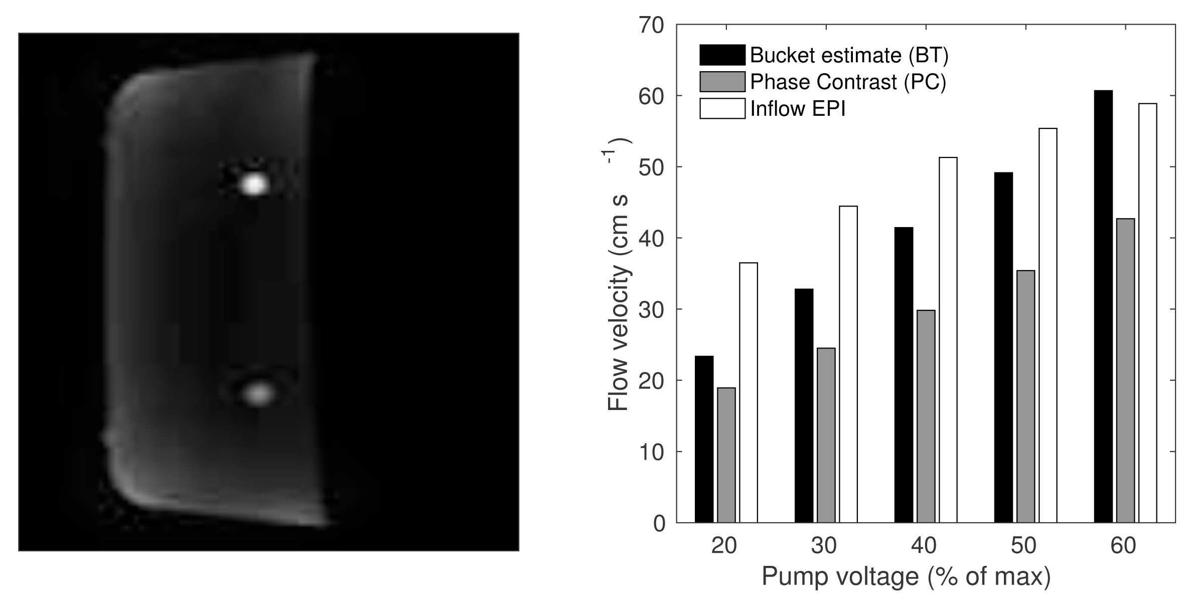
Flow velocity (FV) quantification experiments were performed on a custom-made flow-phantom, pumping a glycerine solution through a 10mm diameter pipe. Varying pump voltage from 10 to 60% of the maximum to modulate the flow velocity, a single EPI slice was used to sample the flow-velocity (256 repeats), and FV was quantified with an estimate of M03. Independent FW estimates were derived using phase contrast (PC) imaging, and from the time taken to fill a litre bucket (BT).
In-vivo experiments
A multi-slice acquisition was used to scan a healthy male subject (4096 repeats, 8 slices, ~8 min scan time). Transverse slices were positioned in the neck and lower head region to cover the Internal Carotid Arteries (ICA). A single sagittal slice scan was also performed at the position of the right Middle Cerebral Artery (MCA). Photoplethysmography (PPG) of the finger was used to measure the cardiac cycle.
Image analysis
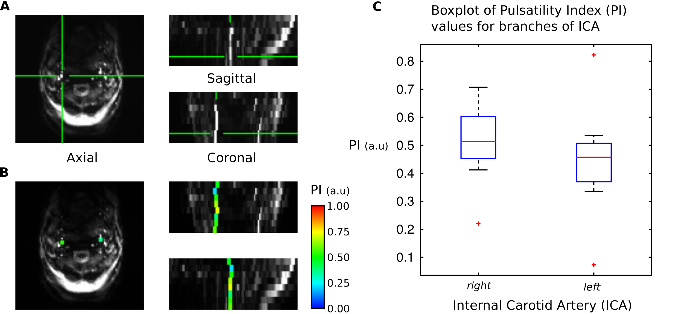
The cardiac phase was discretised into 500 bins in order to rearrange k-space lines to create 500 ‘composite’ k-space planes for each slice, and corresponding series of dynamic images with a temporal resolution of ~2ms (mean cardiac period/500). Vessel mask average time series were obtained for each slice of the ICA and a Fourier series fit generated smooth waveforms from which the Pulsatility Index was calculated (PI). The time between the ‘feet’ of the ICA and MCA waveforms was used to calculate the timing difference, which can be used to estimate PWV by assuming a constant timing offset relative to the PPG.
Results
Phantom experiments
Fig.1 shows the inflow estimates of FV compared with PC and BT. Inflow estimates increase monotonically with pump voltage in agreement with PC and BT estimates. Systematic biases between the techniques need further investigation and may be related to differing sensitivities to vortical flow. Obtaining in-vivo quantitative FV measure in smaller cerebral arteries is currently limited due to partial volume contamination caused by low spatial resolution.
In-vivo experiments
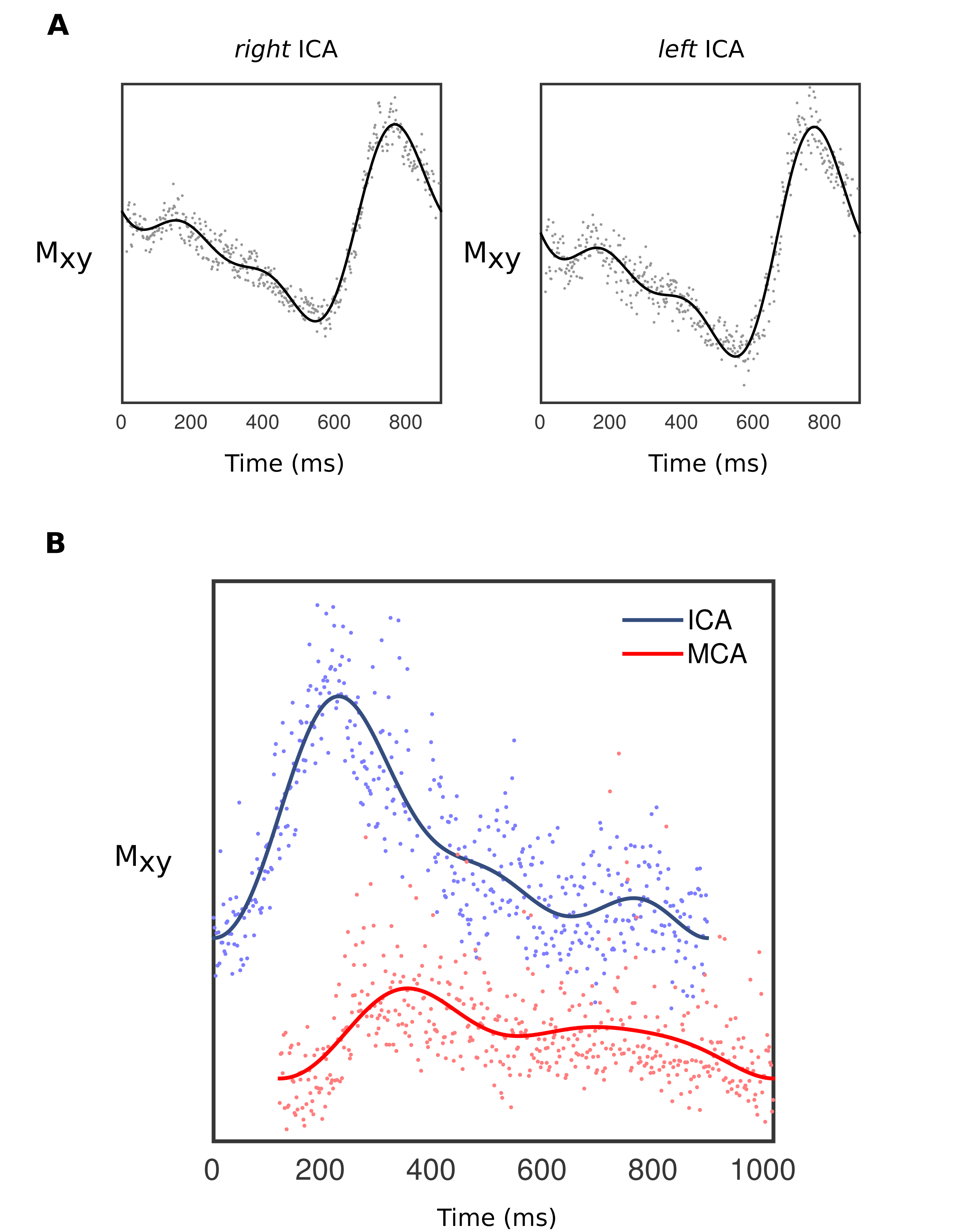
Fig.2 shows cardiac phase ‘composite’ k-space reconstructed images from which the ICA can be seen, along with bilateral PI estimates along slices. Fig.3 shows the slice average waveforms for each branch of the ICA, and the right ICA (slice 1) and MCA waveforms. There is a phase difference of 119.23 ms between ICA and MCA, from which PWV could be quantified if a measure of the streamline distance between the vessel segments was available. Assuming a distance of 30 cm, PWV was calculated to be 2.5 m s-1, which is of the correct order of magnitude.
Discussion
This study demonstrates that fast EPI is sensitive to FV, which when quantified is in relatively good agreement with BT estimates. Retrospective binning of k-space lines allows pulsatile CBFV in cerebral arteries to be measured with high temporal resolution (~2ms, comparable to TCD). With multi-slice acquisitions it's possible to measure waveforms along the arterial tree, allowing markers of AS such as PI to be mapped onto the vasculature.
We show a measurable timing difference in the pulsatile flow waveforms between ICA and MCA, which can be translated into PWV if the distance between the points is known. Waveform shape differences between these two locations likely reflect pressure buffering in the Circle-of-Willis. This is a promising approach for high temporal resolution imaging of CBFV in cerebral arteries (including those inaccessible to TCD) to yields measures of vascular health such as PWV.
Acknowledgements
This work was supported by the Wellcome Trust [WT200804]References
1. Li, X., et al., Arterial stiffness and cognitive impairment. J Neurol Sci, 2017. 380: p. 1-10.
2. Shirwany, N.A. and M.H. Zou, Arterial stiffness: a brief review. Acta Pharmacol Sin, 2010. 31(10): p. 1267-76.
3. Bianciardi, M., et al., The pulsatility volume index: an indicator of cerebrovascular compliance based on fast magnetic resonance imaging of cardiac and respiratory pulsatility. Philos Trans A Math Phys Eng Sci, 2016. 374(2067).
4. Whittaker, J.R., Liebig, P., Fasano, F., Venzi, M., Heidemann, R., and Murphy, K. Cerebrovascular function in the middle cerebral artery measured using the cardiac-induced inflow effect on fast echo-planar imaging. in Proc. Int. Soc. Magn. Reson. Med. 2018. Paris.
Figures