2716
A Simple Homogeneity Correction for Neuroimaging at 7T1High Field Magnetic Resonance Center, Department of Biomedical Imaging and Image-Guided Therapy, Medical University of Vienna, Vienna, Austria
Synopsis
A wide range of MR sequences produce inhomogeneous magnitude images due to the coil sensitivity variation over the head, which is especially severe for ultra-high field strengths. The optimum solution would be a homogeneous reference coil, which however is not possible at 7T due to the shorter wavelength. To date, correction methods require a very long computation time rendering them impractical for on-console imaging. We propose a new magnitude inhomogeneity correction approach, which is based on simplified segmentation and fast interpolation to estimate the bias field. The resulting images show high homogeneity across all three dimensions without any visible artifacts.
Purpose
To improve the homogeneity of MR magnitude images with an approach which is sufficiently computationally simple to allow it to be performed on the scanner reconstructor.Introduction
Inhomogeneity in magnitude images presents as a smooth intensity variation across the head which complicates visual inspection and leads to errors in automated processing such as segmentation. The origin of inhomogeneities is the spatially varying coil sensitivities $$$S_i$$$, where the measured magnitude $$$M_i$$$ can be written as (not considering noise);
$$M_i=S_i*K,$$
in which $$$K$$$ is the true imaged object and $$$i$$$ the coil number.
A common method for the combination of separate-channel magnitude images over a number of coils is root sum-of-squares (RSOS). The magnitude image from each coil serves as an estimate of the coil sensitivity. Squaring the magnitude introduces weighting which yields close-to-optimum SNR but exacerbates inhomogeneity. Taking the square root of the sum-of-squares images partially reduces this1 as can be seen in Fig.1.
$$\text{RSOS}=\sqrt{\sum{M_i^2}}=\sqrt{\sum{(S_i^2*K^2)}}=K\sqrt{\sum{S_i^2}}$$
If the sensitivities of the coils, which change in the presence of the object, are known, sensitivity-weighted combination can be performed:
$$\frac{\sum{(M_i*S_i)}}{\sum{S_i^2}}=\frac{\sum{(K*S_i^2)}}{\sum{S_i^2}}=\frac{K\sum{S_i^2}}{\sum{S_i^2}}=K$$
Several methods (e.g. Damen 20182) estimate the coil-sensitivities to perform sensitivity weighted combination. This is time consuming, however, as it requires the calculation of a smoothed sensitivity profile for each channel, which requires segmentation and interpolation, followed by fitting or smoothing. At high field strengths, the shape of the sensitivities is complicated due to the reduced wavelength and imhomogeneous profiles (Fig.1, sensitivities).
We propose a new homogeneity correction that operates on combined RSOS magnitude images and removes the bias field by estimating and dividing $$$\sqrt{\sum{S_i^2}}$$$ using similar, but simplified steps compared to the separate-channel approach described above.
Methods
The proposed method starts with a simplified segmentation that removes the highest and lowest values from the image in a box-wise approach (Fig.2). A modified moving average filter is then applied iteratively to smooth and interpolate the segmented image to obtain a sensitivity map for the combined image (Fig.3). Finally, the magnitude of each echo is divided by this combined sensitivity map to produce homogeneous magnitude images.
For each dimension, the modified moving average filter was applied 8 times to approximate 3D Gaussian filtering. The size of the individual moving average windows was chosen to obtain an effective sigma of 7mm. The calculation steps are presented in a github repository3.
Data was acquired from a healthy volunteer with a 7T MR whole body Siemens MAGNETOM scanner with a 32-channel Nova Medical head coil. The head was placed slightly off-center to achieve similar inhomogeneities as would be encountered with a larger head.
Scan 1 was 3D monopolar multi-echo gradient echo with a matrix size 416x375x224, TE=[4.5;9]ms, voxel size of 0.5mm, receiver bandwidth of 388 Hertz/pixel and GRAPPA of 2, slice and phase partial Fourier factors of 6/8 and TA=328s.
Scan 2 was a sagittal MPRAGE with matrix size 320x310x208, voxel size 0.75mm, receiver bandwidth 220 Hertz/pixel and TA=280s.
Results
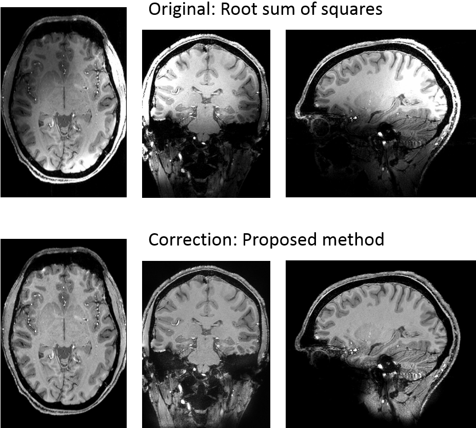
Figs. 4 and 5 show the correction method applied to the gradient echo and MPRAGE scans. The RSOS images show the inhomogeneous intensity which is characteristic at high field strengths. In the corrected images, no visible inhomogeneity is left and no artifacts due to the correction are observed.
The single-threaded prototyped version in MATLAB required 2min 20s to perform the correction of the dual-echo GRE scan of the size 416x375x224.
Discussion
We have presented a simple method for correcting the homogeneity of magnitude images at ultra-high field which produces very homogeneous images without any visible artifacts. The method is heuristic, depending upon the masked tissue being similar throughout the brain, but it nonetheless works for images with very different tissue contrasts such as T1 and T2*-weighted GRE and MPRAGE acquisitions.
In the presence of large tissue regions, the inclusion of SNR-based voxel measures2, but applied only on the combined magnitude could improve the segmentation without sacrificing the fast computation. Another possible improvement would be to perform a phase-corrected and magnitude-weighted complex combination instead of the RSOS method as a first step (as in ASPIRE4 and VRC5) to improve the noise characteristic of the image.
Further work involves the implementation of the correction algorithm on the scanner image reconstruction computer and a more detailed assessment of the robustness in other body parts and in the presence of pathologies.
Conclusion
We have proposed a method to remove the inhomogeneity of magnitude images which produces very good results for healthy brain images in a short computation time and with simple algorithmic requirements that allows it to be transitioned to on-console application.Acknowledgements
This study was supported by funds of the Austrian Science Foundation FWF, Project Number 31452.References
1. Belaroussi B, Milles J et al. Intensity non-uniformity correction in MRI: Existing methods and their validation. Medical Image Analysis 2006; 10:234-246
2. Damen FC and Cai K. $$$B_1^-$$$ non-uniformity correction of phased-array coils without measuring coil sensitivity. Magn Reson Imag 2018; 51:20-283.
3. Eckstein K, Magnitude Intensity Correction, (2018), GitHub repository; https://github.com/korbinian90/Magnitude-Intensity-Correction
4. Eckstein K, Dymerska B et al. Computationally Efficient Combination of Multi-channel Phase Data From Multi-echo Acquisitions (ASPIRE). Magn Reson Med 2018; 79(6):2996-30064.
5. Parker DL, Payne A et al. Phase reconstruction from multiple coil data using a virtual reference coil. Magn Reson Med 2014; 72(2):563-569
Figures
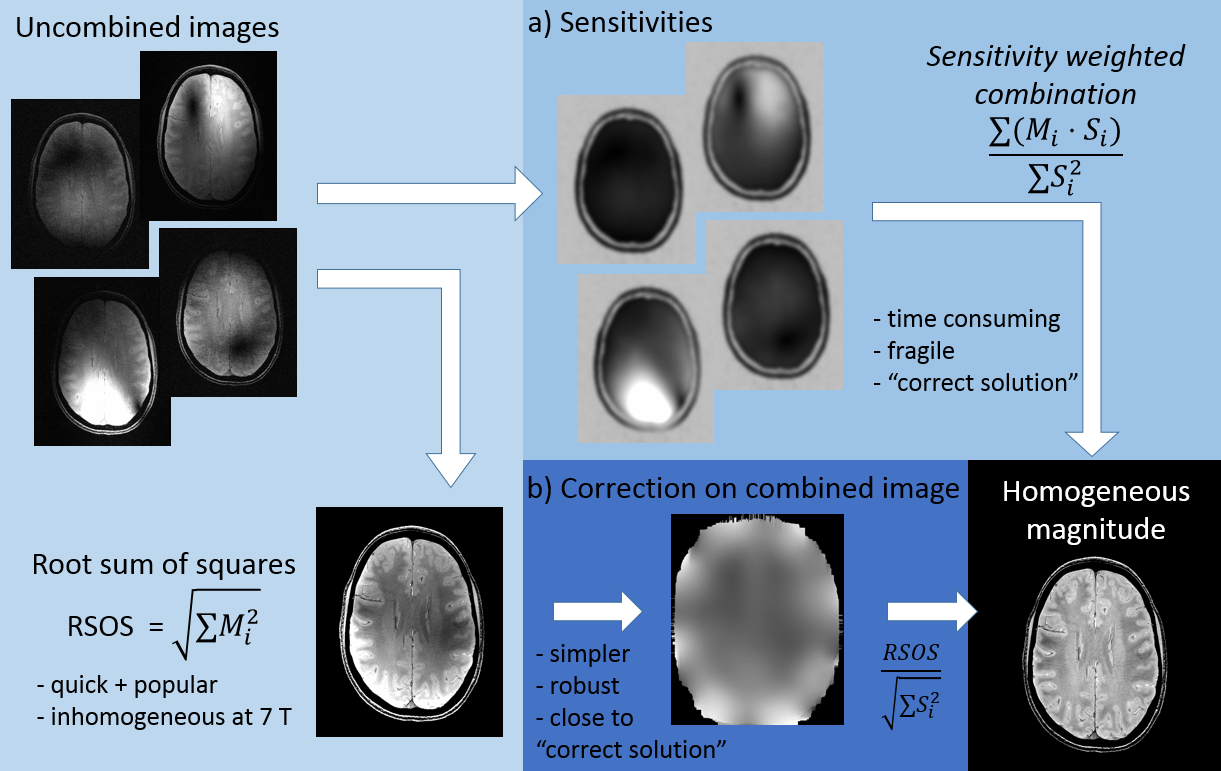
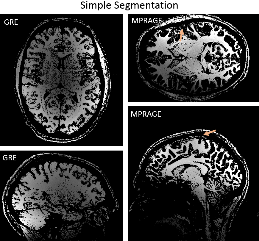
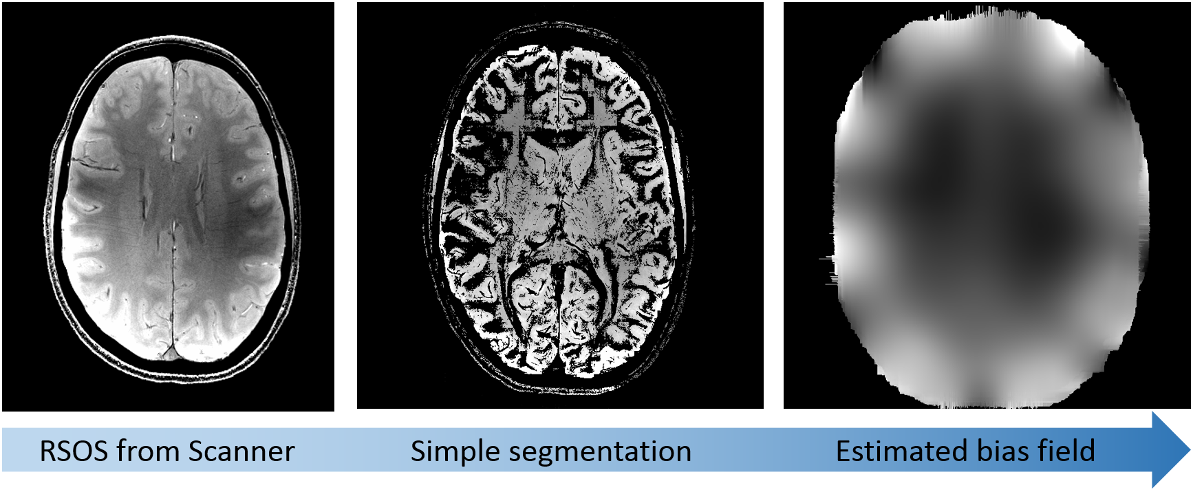
Figure 3: Bias field estimation. The simple segmentation is performed on the RSOS image from the scanner and works well on the inhomogeneous magnitude using an “overlapping box” approach. The segmented image is interpolated and smoothed to obtain an estimation of the bias field √∑Si2. Note that the regular pattern in the estimated bias field corresponds to the position of the coils.
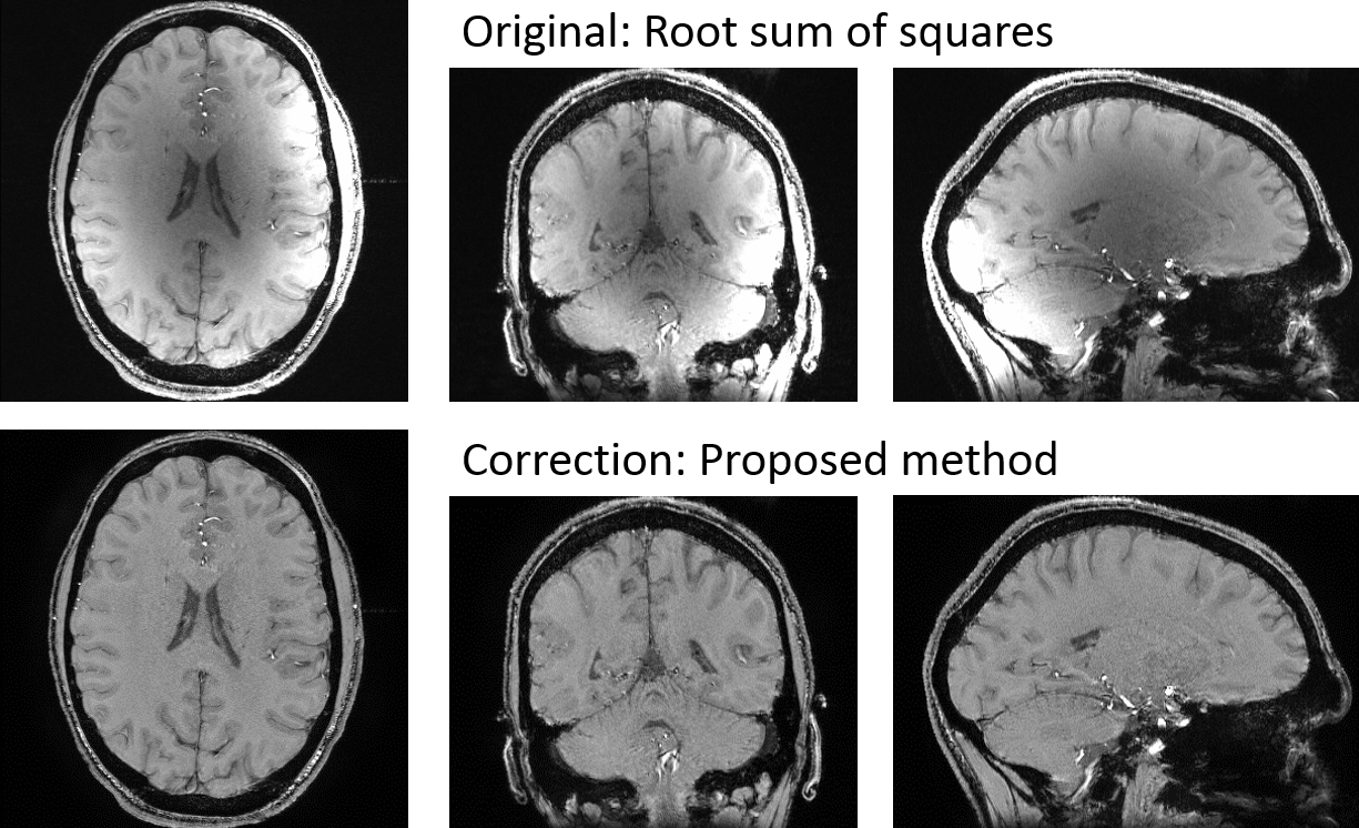
Figure 4: Correction of GRE image. Top row: images combined using RSOS show pronounced inhomogeneity in right posterior and occipital regions. Bottom row: inhomogeneity is effectively removed using the proposed method, which has RSOS images as input. The images have been scaled to the same contrast between grey and white matter.