2714
Automatic segmentation of thalamic nuclei using multiple imaging modalities at ultrahigh field1Translational & Molecular Imaging Institute, Icahn School of Medicine at Mount Sinai, New York, NY, United States, 2Radiology, Icahn School of Medicine at Mount Sinai, New York, NY, United States
Synopsis
Segmenting gray matter structures within the thalamus is complicated by poor inherent T1/T2 contrast. Most existing approaches focus on clustering diffusion data including fiber orientation and short & long distance diffusion directions. We propose a hybrid approach incorporating diffusion data with a recently-developed high T1 contrast imaging sequence known as FGATIR. The proposed algorithm clusters on spatial position, fiber orientation distribution coefficients and anatomical contrast to provide robust, yet fast and fully-automatic segmentation of the thalamic nuclei showing strong agreement to manual segmentation performed by a neuroradiologist. Reliable thalamic nuclei segmentation could facilitate targeted therapies like deep brain stimulation.
Introduction
As the brain’s central relay station, the thalamus plays an essential role in signaling between sensory, motor and associative brain regions. Interest has grown in the role of the thalamus in the assessment of pathologies including epilepsy and multiple sclerosis (MS), and in thalamic nuclei for targeted therapies including deep brain stimulation (DBS). For example, the atrophy of the thalamus has been noted in MS and anterior thalamic nuclei may serve as potential targets for DBS therapies in refractory epilepsy. Reliable magnetic resonance imaging (MRI) based segmentation of thalamic nuclei is challenging due to inherently poor T1/T2 contrast and distinction between thalamic nuclei. Consequently, previous proposals have focused on clustering these structures according to fiber orientation distributions or short and long distance diffusion directions as characterized by diffusion tensor imaging (DTI) [1-5]. We propose a hybrid approach leveraging these diffusion data along with greater anatomical contrast offered by ultrahigh field and a recently-developed imaging technique sensitive to gray matter structures. Considering multiple imaging modalities may provide a robust and specific, yet fast and fully-automatic method for segmentation of the thalamic nuclei.Materials & Methods
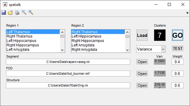
A fast gray matter acquisition T1 inversion recovery (FGATIR) sequence[6] was developed to facilitate increased gray matter contrast with parameters including: T/TR=3.61/3550ms, θ=8°, 409 ms inversion pulse to null white matter, 0.7mm3 isotropic resolution, 11 minute scan time. DTI parameters included: TE/TR=67.6/6000ms, B-value=1500s/mm2, 68 directions, 1.05mm3 isotropic, 18:38 minute scan time. All scans were performed using a 32-channel Nova head coil on a Siemens 7T MAGNETOM scanner, and diffusion & anatomical data from 10 healthy volunteers were assessed for this developmental study. MRTrix was used to process diffusion data using constrained spherical deconvolution to resolve crossing fibers and generate 48 fiber orientation distribution (FOD) coefficients. Segmentation of the thalamus was performed on anatomical imaging data using FreeSurfer 6 and image registration was performed using the Statistical Parametric Mapping (SPM) toolbox in Matlab. A modified K-means clustering algorithm, known as Spatial-K, was developed to segment according to the three spatial dimensions, 48 FOD coefficients and up to two anatomical contrasts (typically T1-weighted anatomical imaging like FGATIR along with susceptibility weighted imaging). The algorithm outputs volume for each of the segmented nuclei and symmetry metrics based on Dice coefficients compared to the contralateral thalamic nuclei. Loop iterations allowed optimization of weighting parameters and number of clusters per thalamus. Loading and segmentation operations could be performed with a core i7 based laptop PC in about one minute per subject. Simultaneously, volumetric manual segmentation of the thalamus from a single high-contrast FGATIR dataset was performed by a neuroradiologist with over twenty years of experience, and the aid of the Morel thalamic atlas. Optimization was performed over 7000 iterations of the algorithm (7x10x10x10 for the available parameters) to compare dice coefficients between the manually and automatically delineated boundaries. Figure 1 shows graphical user interface of the segmentation algorithm.Results
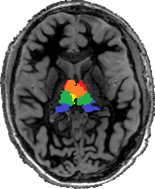
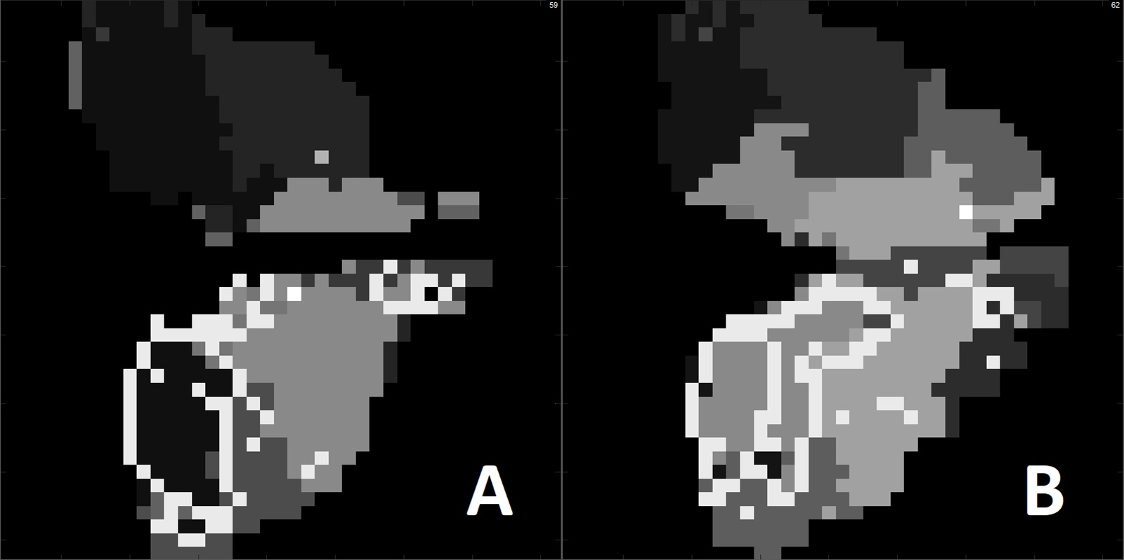
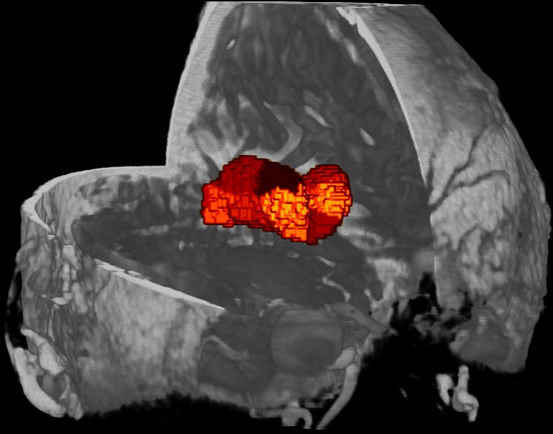
Figure 2 shows the results of thalamic segmentation against an axial FGATIR dataset. Figures 3A and 4B show segmentations for a second subject comparing automatic and manual segmentation at two slice levels. Figure 4 shows a volumetric representation of the thalamic nuclei superimposed on T1-weighted imaging. The algorithm showed good agreement between the two sets of boundaries particularly in nuclei such as the pulvinar. It should be noted that the segmentation algorithm clustered on both anatomical contrast and fiber orientations, whereas manual segmentation was performed solely with anatomical data. Empirical optimization yielded segmentation parameters including: 7-8 clusters weightings of spatial (0.4 relative weighting), diffusion (0.6) and anatomical contrast (0.6) parameters. These approximately-equal weightings also served to justify the consideration of each of the included imaging modalities in the segmentation algorithm. Symmetry measures across the ten healthy controls reflected Dice coefficients of 0.55 to 0.75 compared to the contralateral thalamic nuclei, confirming a pattern of symmetry observed with previously-implemented segmentation techniques.Conclusion
We have demonstrated a fast and reliable technique for thalamic nuclei segmentation using data from multiple imaging modalities and benefitting from the high resolution and contrast of ultrahigh field. Reliable automatic segmentation could have clinical utility in the assessment of pathologies including epilepsy, multiple sclerosis and psychiatric disease. Some thalamic structures, like the anterior nuclei, have been proposed as potential targets for DBS therapy in epilepsy.Acknowledgements
The authors would like to acknowledge the contributions of Ms. Judy Alper and funding from NIH R01 MH109544.References
1. Battistella, G., et al., Robust thalamic nuclei segmentation method based on local diffusion magnetic resonance properties. Brain Structure and Function, 2017. 222(5): p. 2203-2216.
2. Rüsch, N., et al., Prefrontal–thalamic–cerebellar gray matter networks and executive functioning in schizophrenia. Schizophrenia research, 2007. 93(1): p. 79-89.
3. Deoni, S.C., et al., Segmentation of thalamic nuclei using a modified k-means clustering algorithm and high-resolution quantitative magnetic resonance imaging at 1.5 T. Neuroimage, 2007. 34(1): p. 117-126.
4. Ziyan, U., D. Tuch, and C.-F. Westin. Segmentation of thalamic nuclei from DTI using spectral clustering. in International Conference on Medical Image Computing and Computer-Assisted Intervention. 2006. Springer.
5. Wiegell, M.R., et al., Automatic segmentation of thalamic nuclei from diffusion tensor magnetic resonance imaging. NeuroImage, 2003. 19(2): p. 391-401.
6. Sudhyadhom, A., et al., A high resolution and high contrast MRI for differentiation of subcortical structures for DBS targeting: the Fast Gray Matter Acquisition T1 Inversion Recovery (FGATIR). Neuroimage, 2009. 47: p. T44-T52.
Figures