2713
Toblerone: partial volume estimation on the cortical ribbon1Institute of Biomedical Engineering, University of Oxford, Oxford, United Kingdom, 2Wellcome Centre for Integrative Neuroimaging, FMRIB, Nuffield Department of Clinical Neurosciences, University of Oxford, Oxford, United Kingdom, 3Department of Neuroscience, Washington University School of Medicine, St Louis, MO, United States
Synopsis
Toblerone is a new method for estimating partial volumes on the cortical ribbon using surfaces as input (eg those produced by FreeSurfer). Evaluation has been performed using both simulations and subjects drawn from the Human Connectome Project. The estimates returned differ from those produced by existing tools such as FSL's FAST, which will have implications for the analysis of functional imaging data (notably ASL). A preliminary analysis of an ASL dataset has been performed using Toblerone's PV estimates.
Introduction
Partial volume (PV) effects present a significant challenge for functional image analysis, notably for arterial spin labeling (ASL) in which context they are a major source of confound1,2. Correction for PV effects (PVEc) requires estimates of the PVs present within an image, which are conventionally obtained using volumetric methods such as FSL’s FAST3. A novel approach for the cortex is to use a surface segmentation method (eg FreeSurfer4) and then consider the intersection between the surfaces of the cortex and individual voxels. The Toblerone algorithm has been developed to estimate PVs in this manner.Theory
Toblerone is a purely geometric approach to PV estimation that answers the following question: given a surface intersecting a voxel, what is the volume bounded by the surface and the voxel? Estimation is performed by considering the cortical surfaces in turn (both the WM/GM and GM/CSF boundaries) to produce voxel-wise PV estimates for GM, WM and non-brain. The key theoretical step within this framework is a modified form of the ray intersection test traditionally used within computer graphics5 that reduces computational complexity.Methods
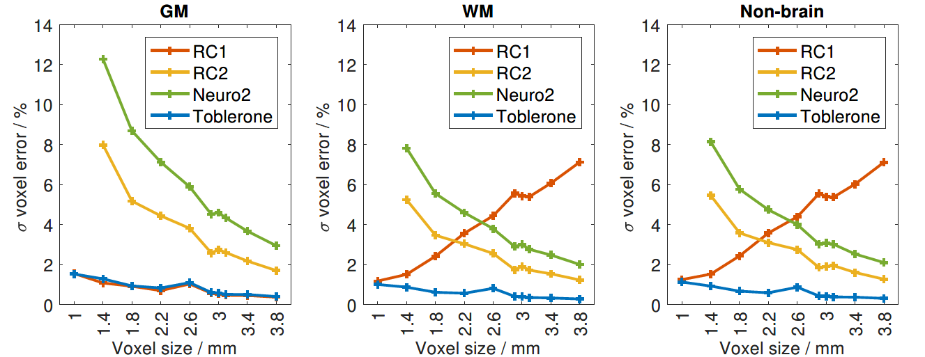
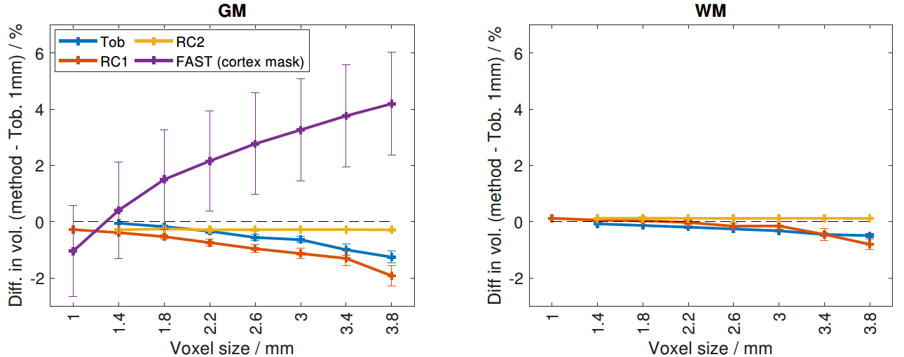
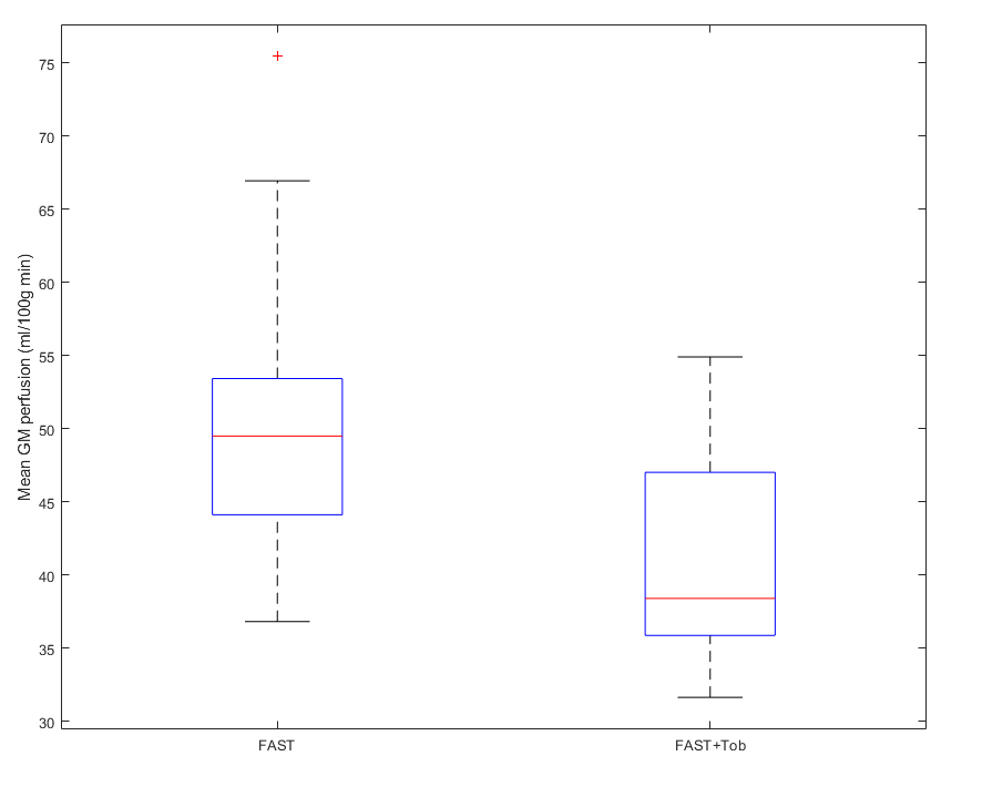
Toblerone has been evaluated using both simulated cortical surfaces and 50 subjects drawn from the Human Connectome Project (HCP). For both types of data, PVs were estimated at voxel resolutions of 1 to 3.8mm isotropic, in steps of 0.4mm. For simulations, errors were calculated on a per-voxel and aggregate basis with reference to ground truth; for HCP subjects, in which context there is no ground truth, total tissue volume was calculated at each resolution to assess within-subject consistency across resolutions. Two other methods were also evaluated on these data (NeuropolyPVE6 and the ribbon-constrained method7), and FAST was also used on the structural scans of the HCP subjects from which their surfaces were produced. Finally, PV estimates produced by Toblerone were used in the analysis of an ASL dataset (multi PLD pcASL, as detailed8, with FreeSurfer as prior step to produce surfaces). As this method produces estimates for the cortex only, subcortical PV estimates were filled in using FSL's FAST. FSL’s oxford_asl pipeline (performing subtraction, calibration, model inversion) was then used to calculate mean GM perfusion in voxels with >80% GM when running on FAST-only or FAST+Toblerone combined PV estimates.
Results
Simulation results showed Toblerone had the lowest, or close to lowest, error of all methods evaluated in both a per-voxel (figure 1) and aggregate sense at all resolutions. HCP results showed good within-subject consistency of total tissue volume across resolutions (figure 3). Results from the task dataset showed lower mean GM perfusion than with FAST-only estimates (fig 4, 41.4 vs 50.1 ml/100g min).Conclusion
Toblerone produces cortical PV estimates with low error (as measured on simulated data) and good consistency (as observed on HCP data) across a wide range of voxel resolutions. These estimates differ to those produced by conventional methods such as FAST and the analysis of an ASL dataset shows that these differences have implications for measured perfusion. Further work is required to determine how Toblerone could be integrated into a wider PVEc framework.Acknowledgements
Funding was provided by the EPSRC (EP/P012361/1) and the Bellhouse scholarship at Magdalen College, Oxford. The authors acknowledge the use of the University of Oxford Advanced Research Computing (ARC) facility in carrying out this work (http://dx.doi.org/10.5281/zenodo.22558).References
1 Zhao M et al. A systematic study of the sensitivity of partial volume correction methods for the quantification of perfusion from pseudo-continuous arterial spin labeling MRI, NeuroImage, 2017
2 Asllani I et al. Regression algorithm correcting for partial volume effects in arterial spin labeling MRI, MRM, 2008
3 Zhang Y et al. Segmentation of brain MR images through a hidden Markov random field model and the expectation-maximization algorithm, IEEE Trans. Med. Imag. 2001.
4 Fischl, B. FreeSurfer, NeuroImage, 2012.
5 Nooruddin et al. Simplification and repair of polygonal models using volumetric techniques, IEEE Trans. Visual. & Comp. Graphics, 2003.
6 Van Assel C et al. Partial volume effect correction for surface-based cortical mapping, Proc. ISMRM, 2017.
7 Glasser M et al. The minimal preprocessing pipelines for the Human Connectome Project, NeuroImage, 2013
8 Mezue M et al. Optimization and reliability of multiple postlabeling delay pseudo-continuous arterial spin labeling during rest and stimulus-induced functional task activation, Jour. Cereb. Blood Flow and Metab. 2014
Figures