2710
Phase-Sensitive Inversion Recovery and T1 Mapping with Motion Correction1Global MR Applications & Workflow, GE Healthcare Canada, Vancouver, BC, Canada, 2Division of Neurology, Department of Pediatrics, BC Children’s Hospital and University of British Columbia, Vancouver, BC, Canada, 3Global MR Applications & Workflow, GE Healthcare, Rochester, MN, United States
Synopsis
In this work, we propose a novel motion corrected Phase Sensitive Inversion Recovery (PSIR) method with integrated T1 mapping derived from MP2RAGE acquisition. Motion correction is achieved using PROMO (PROspective MOtion correction), as well as Optimal Weighted Average (OWA) combination of multichannel data. This proposed method will be useful in obtaining high quality T1 images for children and other subjects who are prone to move during scans.
INTRODUCTION
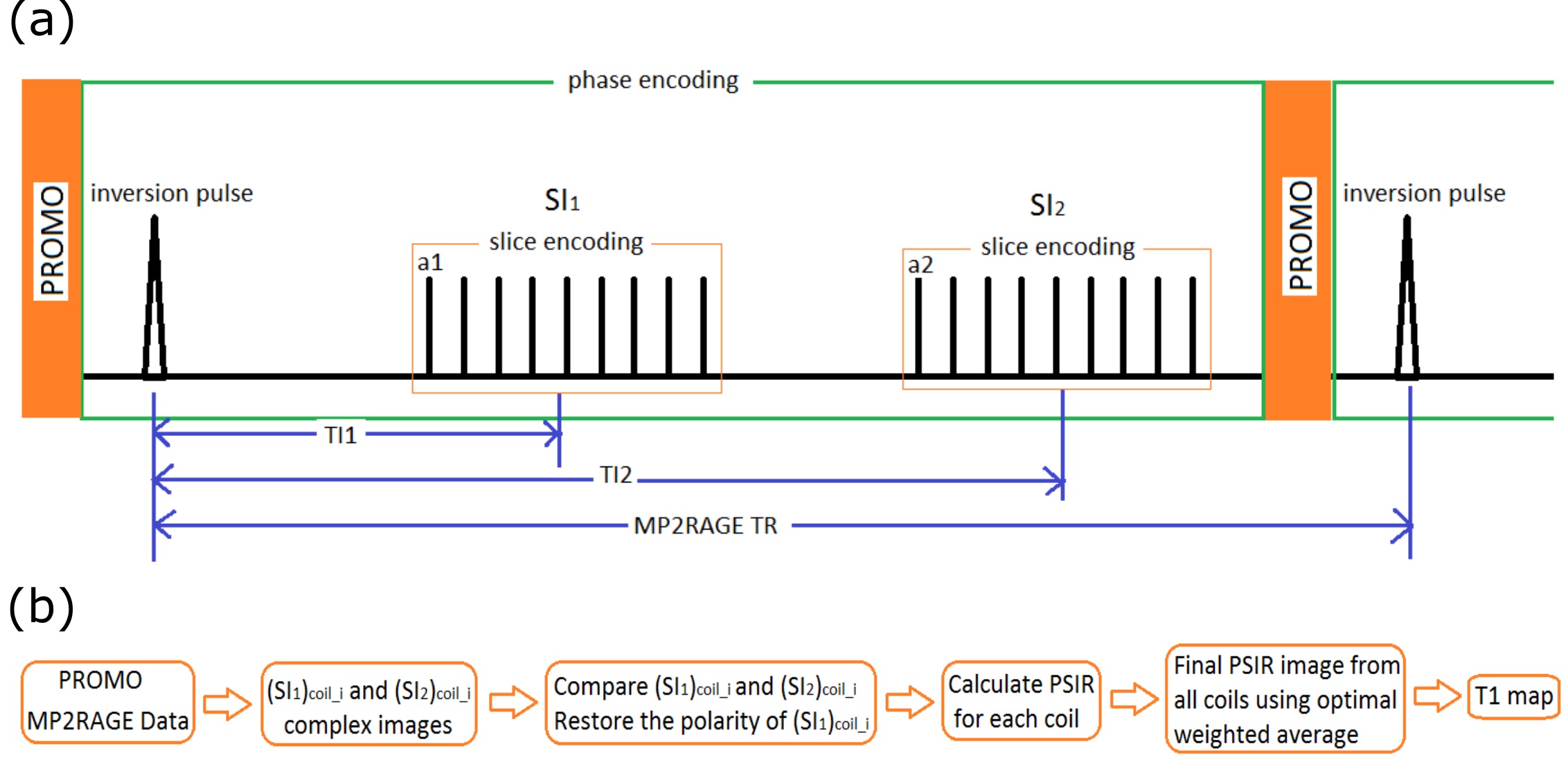
T1 is an essential component of a volumetric brain exam, and motion remains a major cause of image quality failures. The Magnetization-Prepared 2 Rapid Acquisition Gradient Echoes (MP2RAGE) sequence has been introduced to obtain bias-field corrected T1-weighted images with T1 maps at high field(1,2). Recently, a new method was proposed to obtain PSIR images from MP2RAGE sequences(3):
$$$PSIR=\frac{\pm\left|SI_{1}\right|}{\left|SI_{1}\right|+\left|SI_{2}\right|}$$$ [1]
The polarity of $$$\left|SI_{1}\right|$$$ was defined as being negative if the phase change between signal at TI1 ($$$SI_{1}$$$) and signal at TI2 ($$$SI_{2}$$$) lay in the range $$$\pi/2$$$ and $$$3\pi/2$$$.
PROMO is a method to use navigator data to estimate and correct for subject motion in every TR, with little scan time penalty. PROMO significantly reduces motion artifacts, which improves brain image quality for morphometry and cortical surface reconstruction (4,5). OWA has been used to combine multi-channel image to further reduce noise and image degradation due to motion(6)(7). In the proposed work, both PROMO and OWA were used to optimize the image for subjects with motion.
METHODS
All experiments were performed on a 3T GE Discovery MR750 scanner on two healthy volunteers after obtaining informed consent. Two different receive coils were used to evaluate the robustness of the channel combination method.
The first volunteer was scanned using a GE HNS 12-Channel head coil under three conditions: 1) Head motion without PROMO; 2) Head motion with PROMO; 3) No intentional motion. Prior to scan, the subject was trained in a MRI simulator to achieve consistent head-nodding motion upon auditory cue: nod backwards and then return to original position; cues every 1 minute. Approximately 20° of “pitch” rotation was consistently achieved after training. The MP2RAGE pulse sequence (as shown in Figure 1(a)) acquisition parameters were: TRMP2RAGE=5s, TR=5ms, TI1/TI2= 0.7s/2s, $$$\alpha_1=7$$$°, $$$\alpha_2=5$$$°, parallel imaging (ARC) acceleration factor = 2. The acquisition matrix size is 256x128 with 172 sagittal slices, scan duration=6.5 minutes.
The second volunteer was scanned using a GE 32-channel head coil using the same sequence settings except: TR=7.5ms. The acquisition matrix size is 512x266 with 64 axial slices, final reconstructed voxel size = 0.45x0.45x3mm3, scan duration=9 minutes.
The complex images for each coil $$$({SI_1})_{coil_i}$$$ and $$$({SI_2})_{coil_i}$$$ were obtained during the data acquisition. The $$$({PSIR})_{coil_i}$$$ was obtained from Equation [1]. The coil combined PSIR was calculated with OWA. The ‘PSIR versus T1’ plot was calculated based on the equation for signal $$$SI_{1}$$$ and $$$SI_{2}$$$ (1), which is used as a lookup table to obtain T1 maps. Figure 1(b) shows the flowchart of this processing.
RESULTS
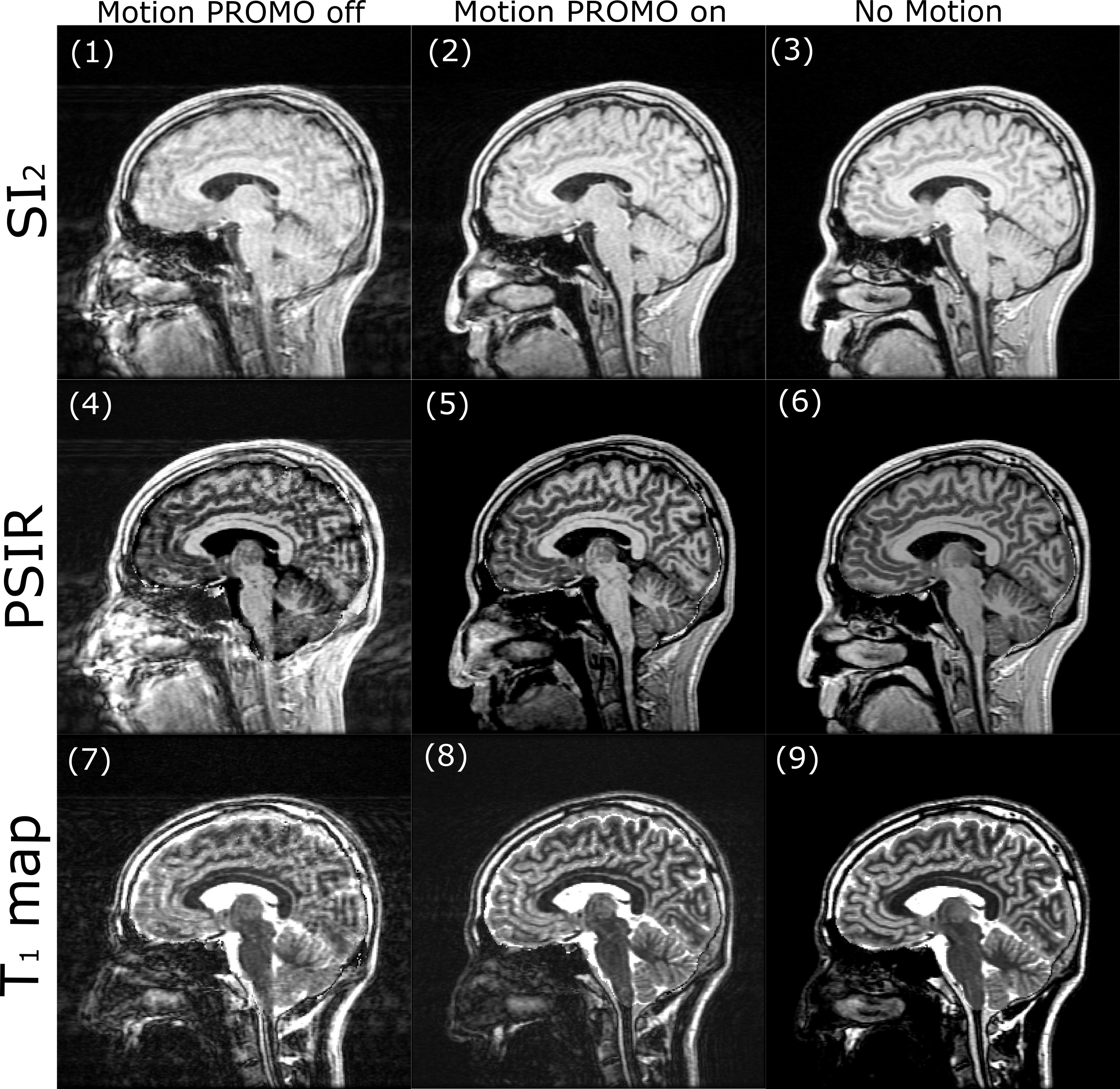
Figure 2 compared the $$$SI_{2}$$$ images, the corresponding PSIR and T1 map for subject with and without motion. As shown in Figure 2(2), with PROMO, motion artifacts can be substantially reduced. However, there is still some residual motion artifact. With OWA, the motion artifact in PSIR images (figure 2(5)) was further reduced. Since the T1 map (Figure 2 (8)) is obtained from the PSIR image, the motion artifact can hardly be detected.
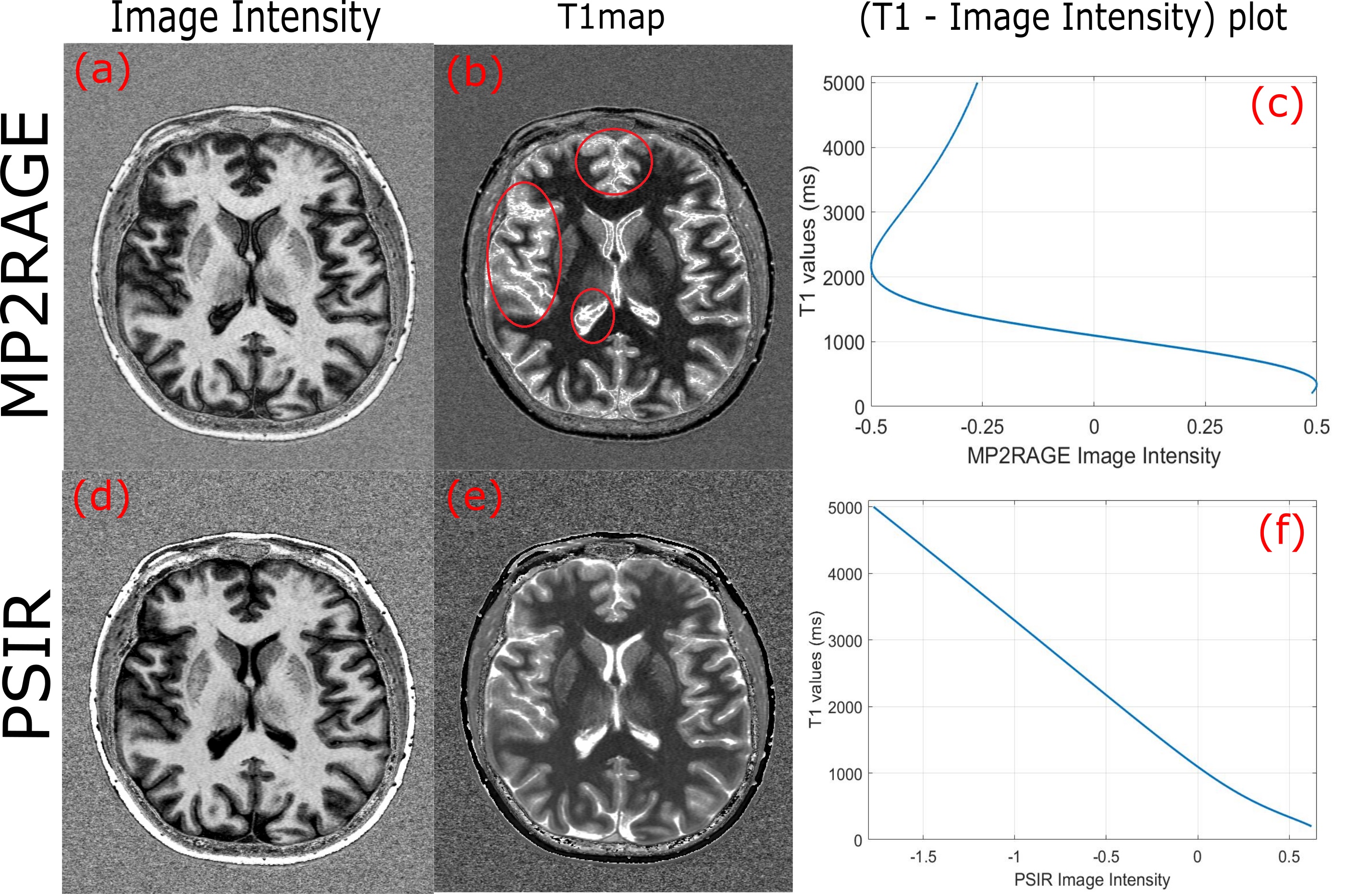
Figure 3 shows a representative high-resolution MP2RAGE (a) and PSIR (d) image. The T1 maps, (b) and (e), were shown correspondingly. The image intensity versus T1 values for MP2RAGE(c) and PSIR(f) were plotted too. As shown in figure3(f), the PSIR signal versus T1 is linear. However, the MP2RAGE signal from approximately -0.5 to -0.25 corresponds to two T1 values (figure3(c)), which causes difficulties in determining some values in the T1 map. The T1 map in figure3 (b) demonstrates areas of some of these intensity discontinuities.
DISCUSSION
From Eq. [1], the PSIR images should be immune to B1 inhomogeneity effect. The PSIR signal fitting can also lead to improved T1 estimation as compared to MP2RAGE method. The T1 maps have no B1 inhomogeneity effect and while an analysis of this was not presented, we believe they will provide an accurate quantitative assessment of tissue T1.CONCLUSION
We have presented a PSIR method derived from a prospectively motion corrected MP2RAGE acquisition utilizing OWA in the multi-channel image combination. The PSIR signal fitting should provide a better estimation of tissue T1 which can be useful for estimation of myelination, and for brain image segmentation and parcellation.Acknowledgements
We thank Michelle Lau for her assistance in data collection. We thank Suchandrima Banerjee for the good comments and suggestions. We also thank everyone in BC Children’s MR Research facility and all the healthy volunteers for the help.References
1. Marques JP, Kober T, Krueger G, van der Zwaag W, Van de Moortele P-F, Gruetter R. MP2RAGE, a self bias-field corrected sequence for improved segmentation and T1-mapping at high field. Neuroimage 2010;49:1271–1281.
2. O’Brien KR, Kober T, Hagmann P, Maeder P, Marques J, Lazeyras F, Krueger G, Roche A. Robust T1-Weighted Structural Brain Imaging and Morphometry at 7T Using MP2RAGE. PLoS One 2014;9:e99676.
3. Mougin O, Abdel-Fahim R, Dineen R, Pitiot A, Evangelou N, Gowland P. Imaging gray matter with concomitant null point imaging from the phase sensitive inversion recovery sequence. Magn Reson Med 2016;76:1512–1516.
4. White N, Roddey C, Shankaranarayanan A, Han E, Rettmann D, Santos J, Kuperman J, Dale A. PROMO: Real-time prospective motion correction in MRI using image-based tracking. Magn Reson Med 2010;63:91–105.
5. Alexandru V. Avram, Joelle E. Sarlls, Cibu P. Thomas, Vinai Roopchansingh, Dan Rettmann, Ajit Shankaranarayanan, Peter J. Basser. Prospective Motion Correction (PROMO) enabled MP2RAGE for multi-contrast high-resolution brain imaging. In: Toronto, Canada; 2015.
6. Taylor JR. Introduction to Error Analysis, Second Edition, John R. Taylor. 2nd ed. University Science Books; 1997.
7. Zhang J, Bjornson B, Xiang Q-S. Combining Multi-channel MP2RAGE Images with Minimized Noise. In: ISMRM 24th Annual Meeting. Singapore; p. #1917.
Figures