2708
A comparison of structural and diffusion-based MRI thalamus segmentation methods1Department of Electrical and Computer Engineering, University of Arizona, Tucson, AZ, United States, 2Department of Psychology, University of California, Los Angeles, Los Angeles, CA, United States, 3Department of Neurosurgery Brain Research Center, University of California, Los Angeles, Los Angeles, CA, United States, 4Department of Medical Imaging, University of Arizona, Tucson, AZ, United States
Synopsis
Automatic thalamus segmentation is a field of study with rapidly evolving applications. Both structural and diffusion weighted MRI can be used to drive parcellations of thalamus nuclei. In this study we present a comparison of leading structural and DWI-based segmentation techniques as implemented on a common set of subject datasets. Results for each are compared, both against an established anatomical atlas and each other. Spatial consistency of nuclei are examined in common template space. Finally, strengths and weaknesses of both techniques are discussed.
Introduction
The thalamus consists of histologically distinct nuclei that serve a variety of functions. These nuclei have been individually implicated in neurological and psychiatric disorders like Parkinson’s disease1 and schizophrenia2. The ventralis intermedius (Vim) nucleus has also been clinically targeted for treatment of essential tremor using deep brain stimulation. MRI-based thalamus parcellation can be used as an aid in volumetry, targeting, and treatment. Most methods developed to date have been based on global or local properties of the diffusion tensor based on an echo-planar imaging acquisition, which is limited in spatial resolution and suffers from distortion. A structural MRI-based thalamic segmentation method has recently been proposed3,4,5 which has been rigorously compared against corresponding manual segmentation with excellent results. This study presents the first direct systematic comparison of structural and DWI-based thalamic segmentation techniques acquired on the same subjects.Methods
The structural segmentation method used for this study was based on a white matter nulled (WMn) MP-RAGE technique using a multi atlas segmentation as described in 3,5. The multi-atlas comprised of 20 prior 7T WMn MP-RAGE datasets with prior manual segmentation using the Morel atlas as a guide. These were registered and averaged to create a mean template. After nonlinear registration to the priors via the template, the 20 sets of labels were fused using PICSL-MALF algorithm 6 to generate a single set of 12 labels representing 10 nuclei, the mammillo-thalamic tract and the whole thalamus. A modified fast version of the algorithm described by Battistella et al 7 was employed to perform DWI-based segmentation. First, the WMn template was registered to the first b=0 volume of the input DWI and used to mask off the thalamus ROI. Orientation distribution functions (ODFs) were fitted to the diffusion signal at each voxel. K-means clustering with a modified distance metric incorporating both Euclidean voxel and ODF coefficient distances was used to generate nucleus labels.Results
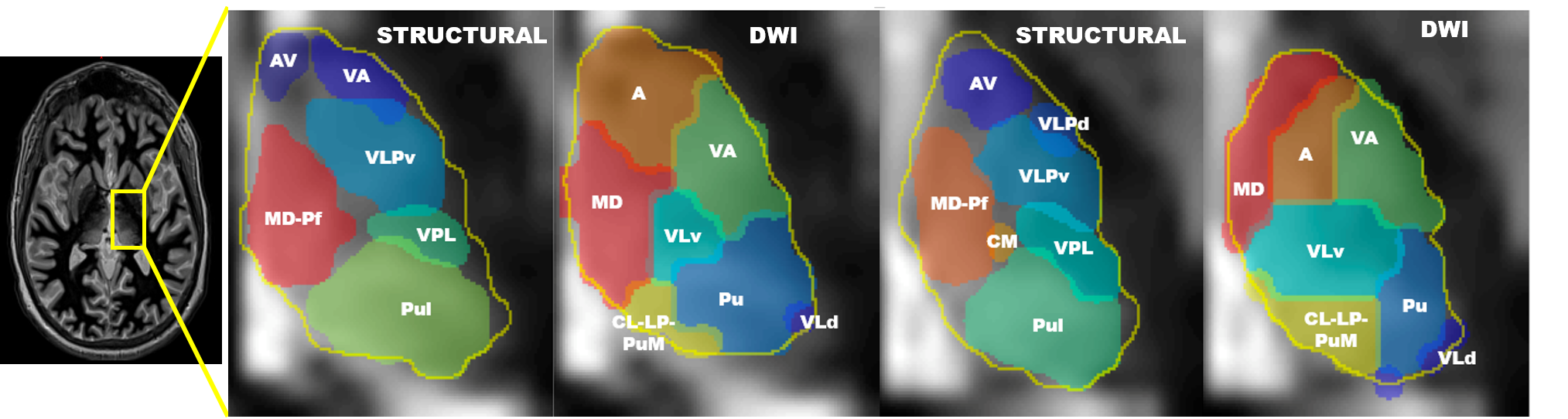
WMn MP-RAGE, DWI, and T1-weighted images were collected for 18 healthy individuals on a 3T Siemens MRI scanned after prior informed consent. Both structural and DWI-based segmentations were run for each case. Figure 1 compares outputs for two of these cases on the same axial slice. Qualitatively, the structural labels tend to correlate more closely with true anatomical structure, while the DWI segmentation tends to produce more spherical clusters, a likely artifact of the k-means algorithm. Evident from the figure is the higher spatial variability of the nuclei generated by the DWI segmentation presumably due to differing local diffusion behavior across subjects.
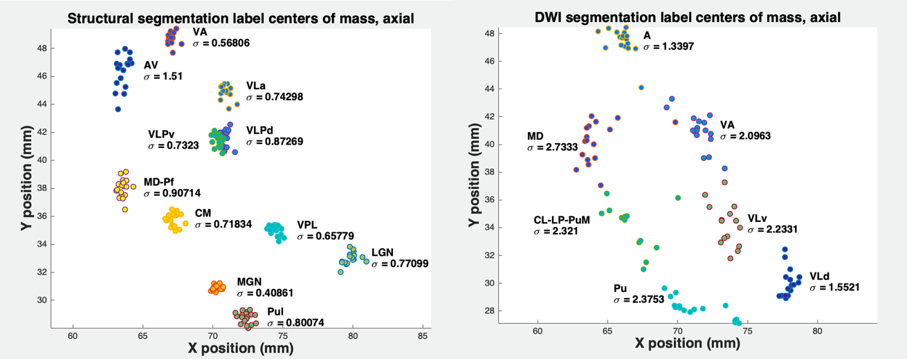
To evaluate the inter-subject consistency, both sets of labels were warped to the WMn template space, and the centroids of those warped labels computed. Figure 2 quantitatively reinforces the observation that the structural labels are more tightly clustered in template space than the DWI labels. Only one set of structural label centroids has a total standard deviation greater than 1.0 mm; none of the DWI-based label centroids have a standard deviation less than 1.0 mm. The structural labels were also warped to the individual DWI space in order to compare the similarities between the two segmentations. Figure 3 shows mean Dice scores between the two methods across all subjects. The measures of coincidence were highest in the mediodorsal (MD), pulvinar (Pul), and the ventral-anterior (VA) nuclei; the similarity scores were lower in the anterior and ventral lateral region.
Conclusion
We presented rigorous comparison of structural and DTI based thalamic segmentation methods on 18 subjects. There are tradeoffs associated with both methods. The WMn-based method produces results which are more consistent with anatomy. It is capable of identifying smaller nuclei such as the lateral and medial geniculate nuclei and the mammillo-thalamic tract. However, it requires a WMn MP-RAGE acquisition which is not part of standard clinical protocols. Conversely, DWI is commonly collected, but image resolution limitations (~2 mm) dictate that DWI-based methods cannot distinguish smaller nuclei which may only span a few voxels. Also, the use of Euclidean voxel distance in the DWI method’s k-means clustering routine tends to bias parcellation outputs towards spherical clusters which are not reflective of thalamic histology. Future research will investigate a fusion of these methods to leverage each other’s strengths. Segmentation could be improved by using local diffusion characteristics to inform structural segmentation, and vice versa.Acknowledgements
No acknowledgement found.References
1. Aggleton et al. "Thalamic pathology and memory loss in early Alzheimer’s disease: moving the focus from the medial temporal lobe to Papez circuit." Brain 139.7 (2016): 1877-1890.
2. Kim et al. Volumetric abnormalities in connectivity-based subregions of the thalamus in patients with chronic schizophrenia. Schizophr Res. 2007 Dec; 97(1-3):226-35. Epub 2007 Oct 30.
3. Su et al. THOMAS: Thalamus Optimized Multi-Atlas Segmentation at 3T. Proceedings of ISMRM. 2016 May 7;4328."
4. Tourdias et al. Visualization of intra-thalamic nuclei with optimized white-matter-nulled MPRAGE at 7T. NeuroImage. 2014;84:534-45.Neuroimage 84 (2014): 534- 545.
5. Thomas et al. A method for near realtime automated segmentation of thalamic nuclei. Proc. of the 25th scientific meeting of the ISMRMl Hawaii 2017, p4736
6. Wang et al. Multiatlas segmentation with joint label fusion, IEEE Trans. Pattern Anal. Mach. Intell., vol. 35, no. 3, pp. 611-623, Mar. 2013
7. Battistella et al. Robust thalamic nuclei segmentation method based on local diffusion magnetic resonance properties. Brain Structure and Function 222.5 (2017): 2203-2216.
Figures