2707
A novel DWI-based thalamus segmentation method using Constrained Spherical Deconvolution1Department of Electrical and Computer Engineering, University of Arizona, Tucson, AZ, United States, 2Department of Biomedical Engineering, University of Arizona, Tucson, AZ, United States, 3Department of Psychology, University of California, Los Angeles, Los Angeles, CA, United States, 4Department of Neurosurgery Brain Research Center, University of California, Los Angeles, Los Angeles, CA, United States, 5Department of Medical Imaging, University of Arizona, Tucson, AZ, United States
Synopsis
Existing methods to segment the thalamus via diffusion weighted MRI are inhibited by several factors. The largely gray matter composition of the thalamus makes the local diffusion activity indistinct and some of the more successful DWI-based methods require time consuming and computationally expensive cortical parcellation for thalamus masking. This study addresses these limitations by using multi-tissue constrained spherical deconvolution to isolate desired diffusion activity and a novel template based technique for thalamus masking. Segmentation outputs are evaluated and we conclude with a discussion of the method’s advantages over existing techniques.
Introduction
The human thalamus, a bilateral, subcortical gray matter structure that acts as a relay station for many neurological processes, is composed of several histologically and functionally distinct nuclei. These nuclei are relatively indistinct using routine anatomical or clinical imaging, and can therefore be difficult to identify for targeted therapy. A number of Diffusion tensor imaging (DTI) based segmentation methods have been proposed, and more recently, more advanced techniques such as Q-ball imaging1 have been proposed. However, because the thalamus is a combination of gray and white matter, and generally has a low fractional anisotropy, it is difficult to reliably estimate primary diffusion orientations and diffusion orientation distribution functions which these methods rely upon. In this work, we propose a new diffusion-based thalamic segmentation algorithm that modifies existing algorithms to use the recent multi-tissue constrained spherical deconvolution (MT CSD)2 technique in order to separate the signal from gray and white matter structures, and more reliably estimate the diffusion properties of the thalamus.Methods
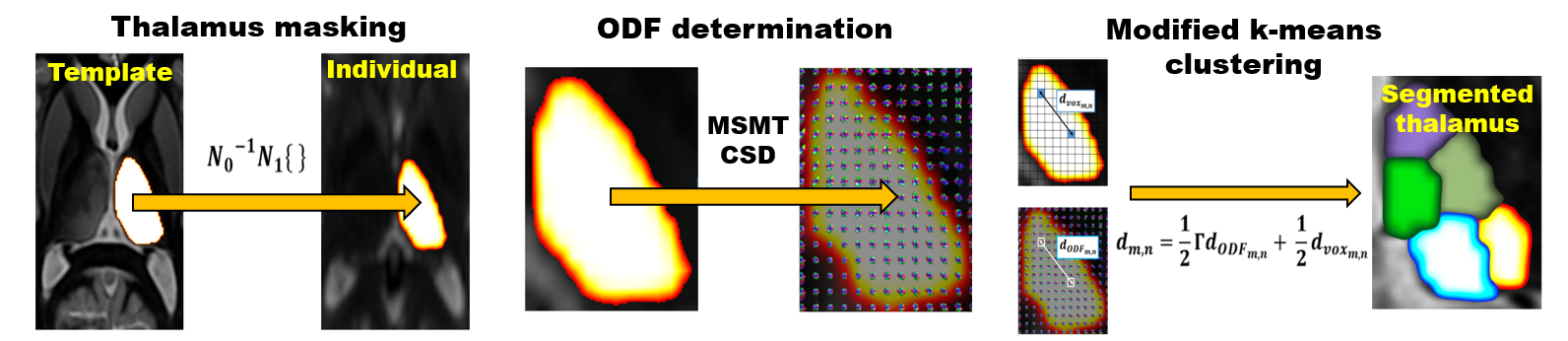
Images were acquired on a 3T Siemens system from 18 healthy subjects after prior informed consent. Diffusion weighted images were collected using a multi-shot EPI sequence (64 directions collected with a =1000 s/mm2, one direction collected with a b-value of 0, voxel size 2mm x 2mm x 3mm). Eddy current and field inhomogeneity distortion correction were performed prior to segmentation. We used a multi-stage registration process to isolate the thalamus. A precomputed thalamic mask from a mean template image was nonlinearly warped to the input space to identify the thalamus ROI. Next, we used MT CSD (with order 6 spherical harmonic basis functions) to generate 28 ODF coefficients. Segmentation was then performed via k-means clustering (N=7 clusters). The distance metric used for k-means was a weighted sum of Euclidean voxel and ODF distances. Output segmentations were evaluated to determine if they corresponded reasonably to anatomical structure. The labels were also transformed to a common template space to gauge spatial consistency.Results
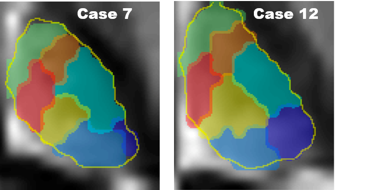
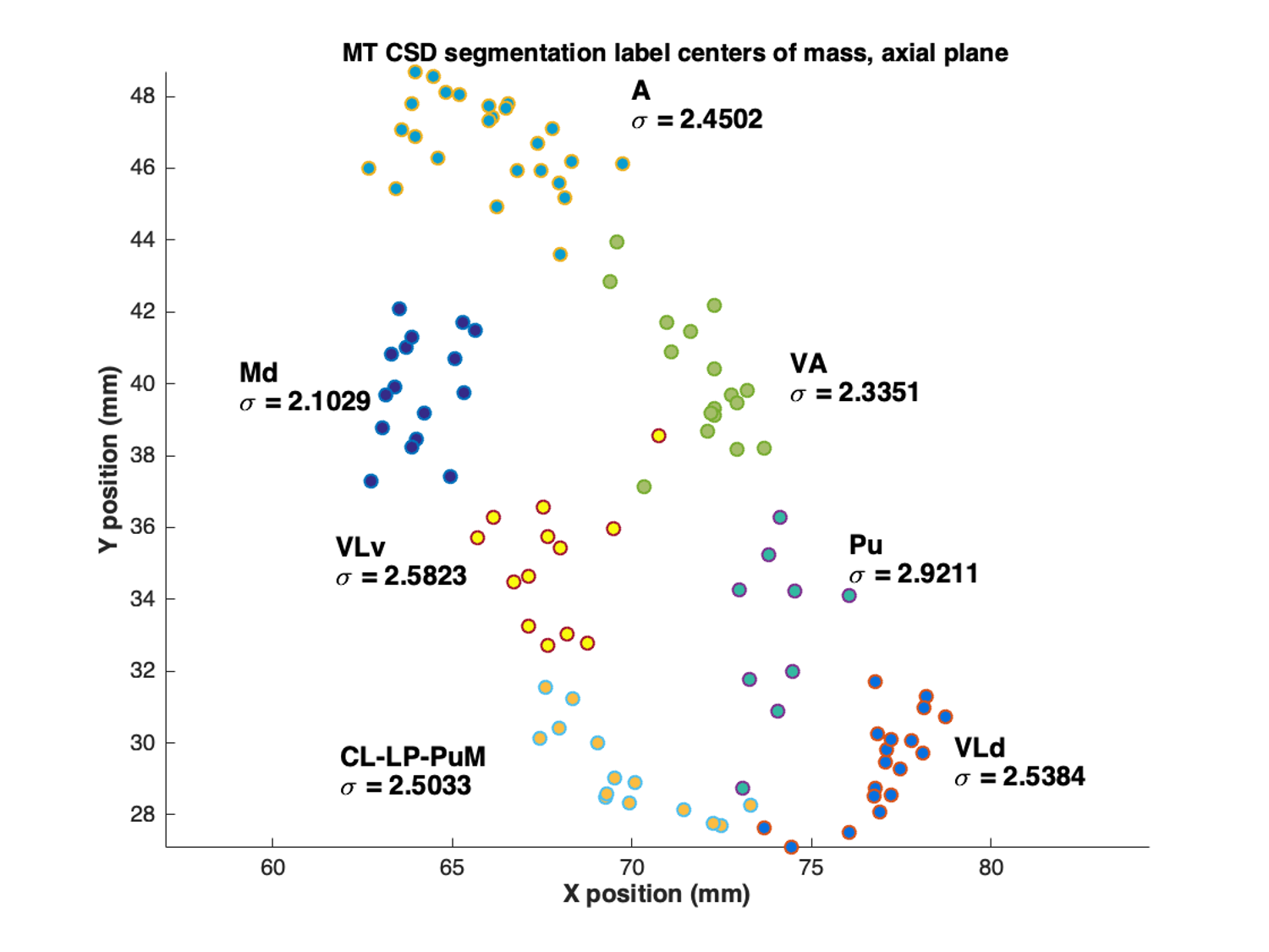
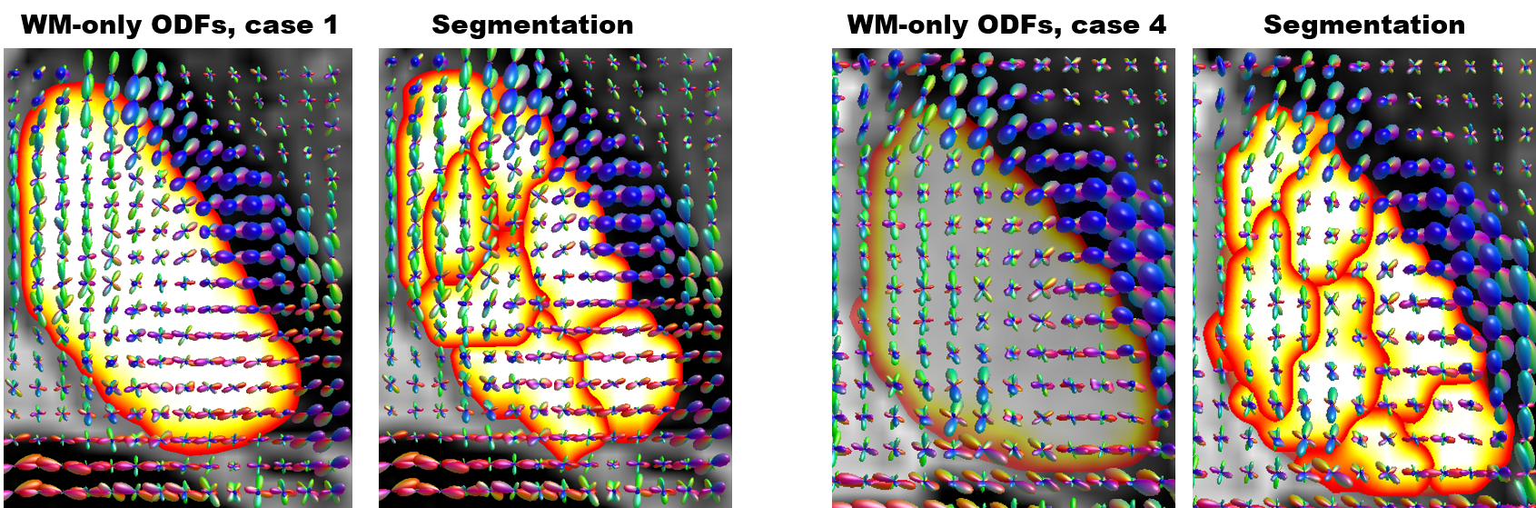
Figure 2 shows two examples of segmentation outputs. The clusters appear to largely correspond with nuclei such as the medio-dorsal and pulvinar. When warped to a common template space, the label centroids exhibit more or less uniform consistency as measured by the spatial standard deviation of the centroid locations (Figure 3). The medio-dorsal centroids have the smallest standard deviation (2.10mm), while the centroids in the pulvinar region have the largest (2.92 mm). As figure 4 shows, the labels appear to exhibit idiosyncratic ODF patterns that are not typically observed in methods involving Q-ball imaging.Discussion
MT CSD has several advantages over Q-ball imaging: 1) It is more robust at smaller b-values (such as the b=1000 s/mm2 used in this study); 2) It disambiguates the diffusion signal as a superposition of white matter, gray matter, and CSF. The largely isotropic tissue responses of gray matter and CSF can then be excluded, with segmentation performed exclusively upon the anisotropic white matter ODFs. As seen in figure 4, the white matter ODFs present a coherent structure that informs the segmentation; 3) It produces sharper ODF peaks; 4) MT CSD executes in seconds when restricted to the thalamus ROI. Also contributing to the overall time savings of this method is the manner in which the entire thalamus is masked off from surrounding tissues. Previous methods2 use time consuming and computationally intensive cortical and subcortical parcellation in Freesurfer and SPM-8 to achieve this (on the order of hours), while we have used a template-based registration technique via ANTs to reach the same result (on the order of minutes).Acknowledgements
No acknowledgement found.References
1. Battistella et al. Robust thalamic nuclei segmentation method based on local diffusion magnetic resonance properties. Brain Structure and Function 222.5 (2017): 2203-2216.
2. Tournier et al. Resolving crossing fibres using constrained spherical deconvolution: validation using diffusion-weighted imaging phantom data. Neuroimage 15: 617-625 (2008)
Figures