2701
A Surface-Constrained Dynamic Elasticity Model for Deformable Registration of Infant Brain MRI1Department of Radiology and Biomedical Research Imaging Center, University of North Carolina, Chapel Hill, NC, United States
Synopsis
Spatial registration of infant brain images is challenging owing to significant changes in image appearance in association with rapid growth in the first year of life. In this abstract, we introduce a volumetric registration method that is constrained by cortical correspondences for consistent cortical and sub-cortical alignment.
Introduction
The infant brain undergoes rapid changes in morphology and appearance during the first year of life. Understanding brain evolution during infancy is substantially important in neuroscience for identifying development disorders. Image registration is a key step for automatic and accurate quantification of brain changes. However, the infant brain image registration is challenging due to the rapid anatomical changes and low tissue contrast between white matter (WM) and gray matter (GM). As a remedy, we propose a surface-constrained volumetric registration method for infant brain MRI.Materials and Methods
The dataset comprised of T1-weighted and T2-weighted scans from $$$19$$$ healthy infant subjects (M: 14, F: 5) enrolled in the Multi-visit Advanced Pediatric (MAP) Brain Imaging Study. Each T1-weighted scan had $$$144$$$ sagittal slices with $$$1.0$$$ mm isotropic voxel size. And T2-weighted scans had 64 sagittal slices with $$$1.25\times1.25\times1.95$$$ $$$\mathrm{mm^{3}}$$$ voxel size. All the scans were processed by the UNC infant cortical surface pipeline1 to generate accurate tissue segmentation maps as well as inner and outer cortical surfaces. To deal with longitudinal appearance changes, we use tissue segmentation maps, instead of intensity images, for registration. We first established the cortical surface correspondence between the fixed and moving subjects using Spherical Demons2. This vertex-wise transformation $$$\Phi(\vec{p_{s}})$$$ is used to constrain the subsequent volumetric registration step, which seeks an optimal transformation $$$\phi(\vec{p_{v}})$$$ such that $$$\phi(\vec{p_{s}})=\Phi(\vec{p_{s}})$$$, where $$$\vec{p_{v}}$$$ and $$$\vec{p_{s}}$$$ define the voxel coordinates and cortical surface vertices, respectively. The underlying large non-linear deformations are modeled using a dynamic elasticity model (DEM) with surface constraints imposed as follows: $$\frac{\partial^{2}{\phi\left(\vec{p_{v}}\right)}}{\partial{t^{2}}}=\alpha\big(\nabla^{2}\phi\left(\vec{p_{v}}\right)+\nabla(\nabla.\phi\left(\vec{p_{v}}\right))\big)+\beta f^{v}\!\left(\vec{p_{v}}\right)+\gamma f^{s}\!\left(\vec{p_{s}}\right),$$
where $$$f^{v}\!\left(\vec{p_{v}}\right)$$$ measures the discrepancy between the fixed and warped moving tissue segmentation maps and $$$f^{s}\!\left(\vec{p_{v}}\right)$$$ gives the error between $$$\Phi(\vec{p_{s}})$$$ and $$$\phi(\vec{p_{s}})$$$. $$$\alpha$$$, $$$\beta$$$ and $$$\gamma$$$ control the data irregularity, data mismatch and deformation error, respectively. We hereby denote our method as SC-DEM.
Results
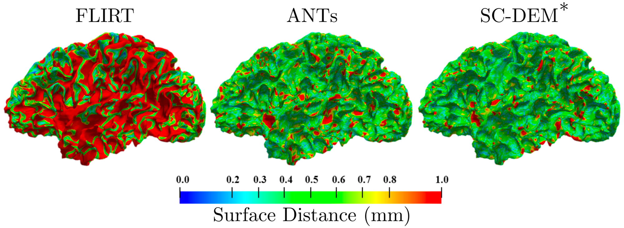
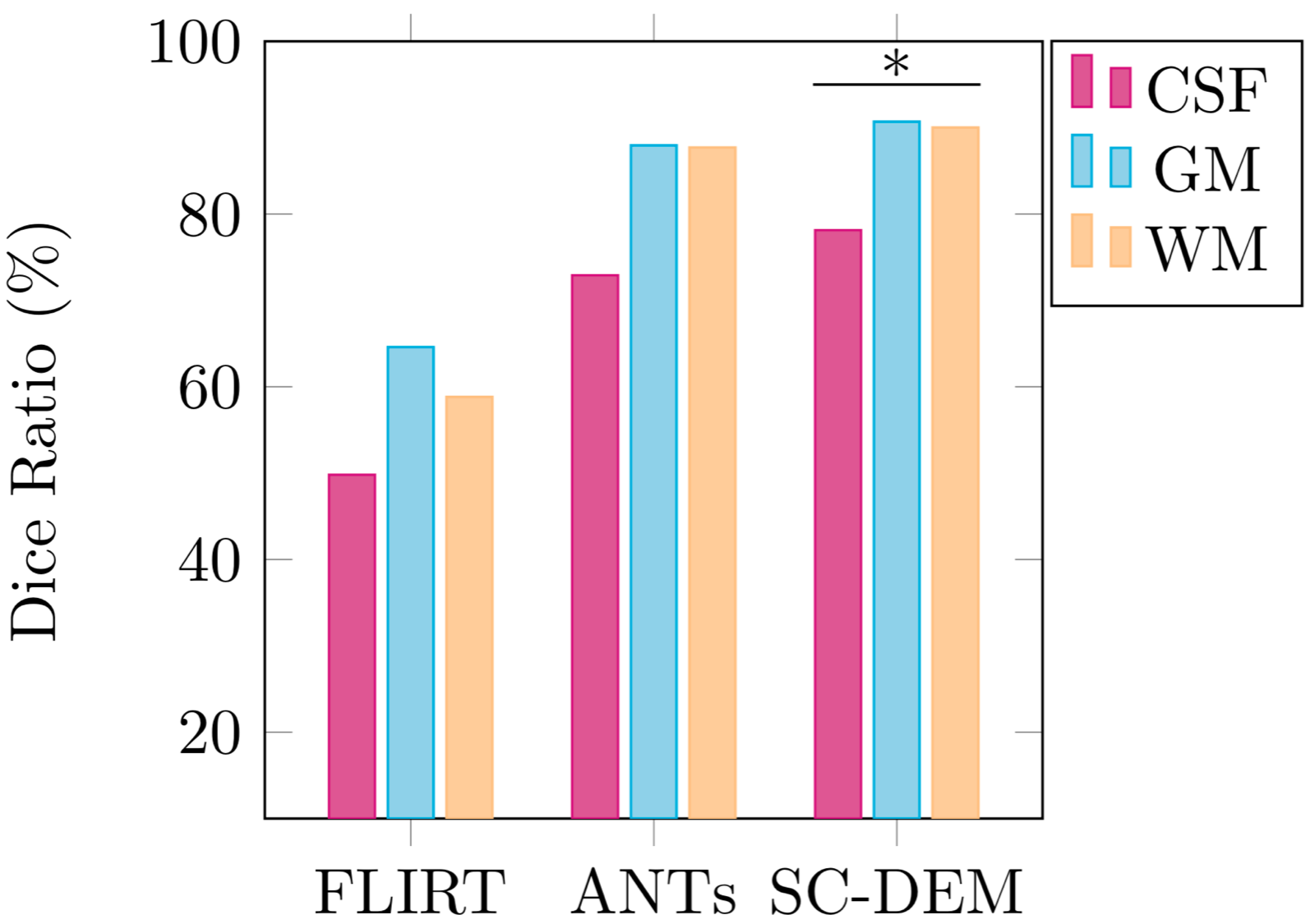
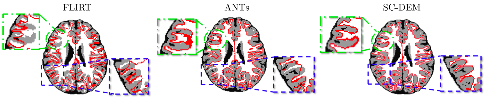
We performed longitudinal registration experiment with 2-weeks-old moving tissue segmentation maps and 12-months-old fixed tissue segmentation maps using FLIRT3, ANTs4 and SC-DEM. The results in terms of cortical surface distance and mean dice ratio are shown in Fig. 1 and Fig. 2, respectively. It can be observed that SC-DEM achieves higher registration accuracy ("$$$^{*}$$$" marks statistical significance with $$$p<0.01$$$) than other methods. Fig. 3 demonstrates the superiority of SC-DEM in cortical alignment.Conclusion
Taking into consideration both surface and volumetric information results in greater accuracy in infant MRI registration.Acknowledgements
This work was supported in part by NIH grants (NS093842, EB022880, EB006733, EB009634, AG041721, and MH100217).References
1. Li G, Nie J, Wang L, Shi F, et al. Measuring the dynamic longitudinal cortex development in infants by reconstruction of temporally consistent cortical surfaces. Neuroimage. 2014; 90:266 - 279.
2. Yeo B T T, Sabuncu M R, et al. Spherical demons: Fast diffeomorphic landmark-free surface registration. IEEE Trans Med Imaging. 2010; 29(3): 650 - 668.
3. Jenkinson M, Smith, S. A global optimisation method for robust affine registration of brain images. Med Image Anal. 2001; 5(2): 143 - 156.
4. Avants B, Epstein C, et al. Symmetric diffeomorphic image registration with cross-correlation: Evaluating automated labeling of elderly and neurodegenerative brain. Med Image Anal. 2008; 12(1): 26 - 41.
Figures