2698
Functional and Structural Abnormality in Patients with Alcohol Use Disorder Combined VBM and FC Analysis1Department of Radiology, Renmin Hospital of Wuhan University, Wuhan, China
Synopsis
We combined voxel-based morphometry (VBM) and seed-based functional connectivity (FC) analysis to identify functional and structural characteristics in patients with alcohol use disorder using high resolution T1-weighted structure images and functional MRI. AUD group showed significantly decreased gray matter volume mainly in the default mode network, and decreased FC in the default mode network and executive control network when compared with the HC group. Combining VBM and FC provides a new perspective on the pathophysiological and clinical manifestations in AUD patients.
Abstract
Introduction Alcohol use disorder (AUD) is associated with numerous structural and functional brain damage[1-3]. Most previous research have focused upon the impact of specific neurological abnormalities. It is clear that the integrated understanding of the brain damage in AUD patients is incomplete. In this study, multi-modal MRI combined with voxel-based morphometry (VBM) and seed-based functional connectivity (FC) analysis were used to investigate functional and structural characteristics in patients with AUD.
Methods Twenty-one AUD individuals and twenty-one age-, sex-, education- and handedness-matched healthy controls were recruited. Cognitive function of all participants were evaluated with the Mini-mental state examination; furthermore, the levels of alcohol-related problems were assessed by the Michigan alcoholism screening test and Alcohol drinking scale (ADS) in AUD group. All the subjects underwent high resolution T1-weighted structure images and functional MRI data acquisition by MR scanning. VBM was used to detect the gray matter volume (GMV), then the brain region with significant group difference in VBM (right cerebellum crus I) was selected as the region of interest to calculate FC. Independent sample t test was performed to evaluate the significant difference of GMV and FC parameters of two groups. Finally, the correlation analysis was performed between GMV/FC in brain regions with significant group differences and clinical variables.
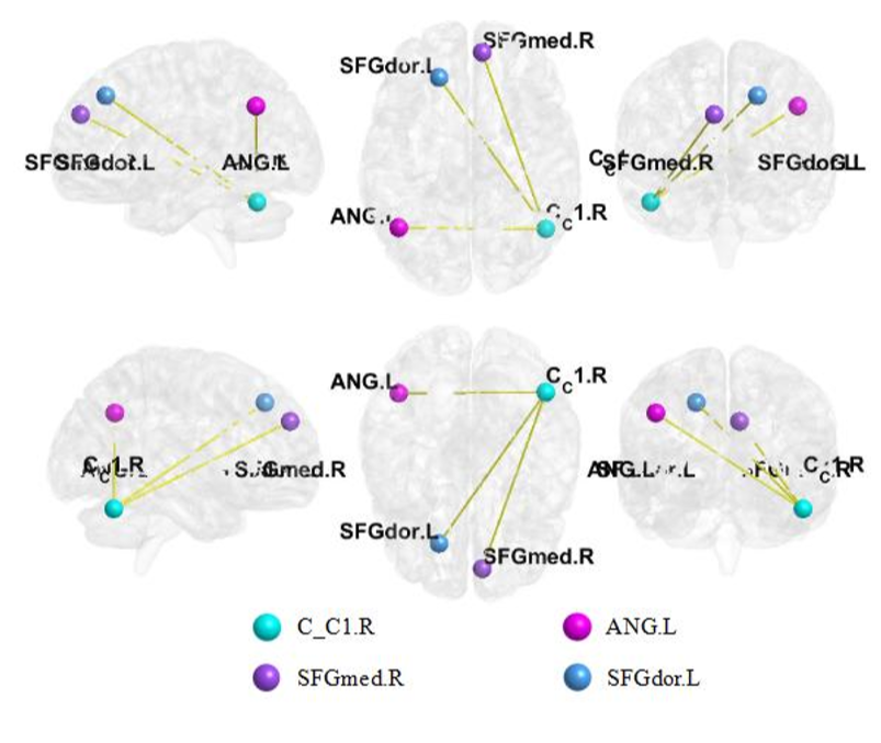
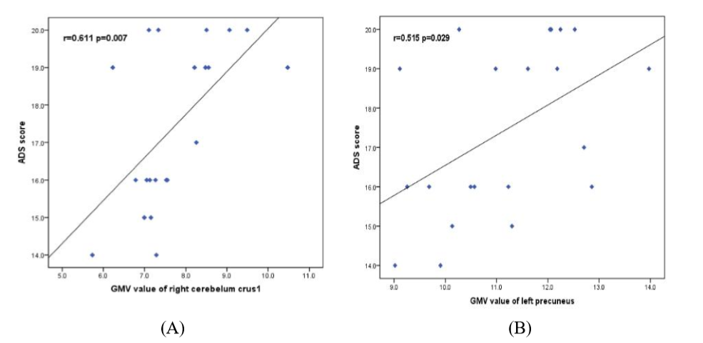
Results Compare with healthy controls, AUD group showed significantly decreased GMV in the left precuneus, right angular gyrus, right postcentral gyrus, right middle temporal gyrus, left cerebellum crus II and right cerebellum crus I (P<0.05, GRF corrected) (Table.1, Fig.1). The AUD group showed significantly decreased FC between the right cerebellum crus I and the right medial superior frontal gyrus, left dorsolateral superior frontal gyrus, and left angular gyrus when compared to healthy controls (P<0.05, GRF corrected) (Table.2, Fig.2). Moreover, in the AUD group, the ADS scores showed a positive correlation with the mean gray matter volume values of the left cerebellum crus 1 (r=0.611; P=0.007) and the left precuneus (r=0.515; P=0.029) (Fig.3).
Conclusion AUD patients exhibited functional and structural abnormality in default mode network and executive control network. Combining VBM and FC provides a new perspective on the pathophysiological and clinical manifestations in AUD patients.
Acknowledgements
At the point of finishing this paper, I’d like to express my sincere thanks to my tutor who have lent me hands.References
1. Yang X, Tian F, Zhang H, Zeng J, Chen T, Wang S, et al. Cortical and subcortical gray matter shrinkage in alcohol-use disorders: a voxel-based meta-analysis. Neurosci Biobehav Rev. 2016; 66:92-103.
2. Jansen JM, van Wingen G, van den Brink W, Goudriaan AE. Resting state connectivity in alcohol dependent patients and the effect of repetitive transcranial magnetic stimulation. Eur Neuropsychopharmacol. 2015; 25(12):2230-2239.
3. Weiland BJ, Sabbineni A, Calhoun VD, Welsh RC, Bryan AD, Jung RE, et al. Reduced left executive control network functional connectivity is associated with alcohol use disorders. Alcohol Clin Exp Res. 2014; 38(9):2445-2453.
Figures
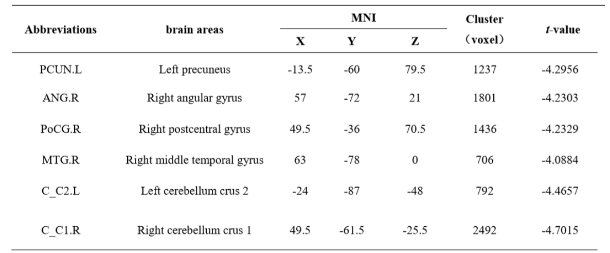
TABLE 1 | Brain areas with decreased GMV values in the AUD group compared with the HC group. Abbreviations: AUD, alcohol use disorder; HC, healthy controls; GMV, gray matter volume; MNI, montreal neurological institute.
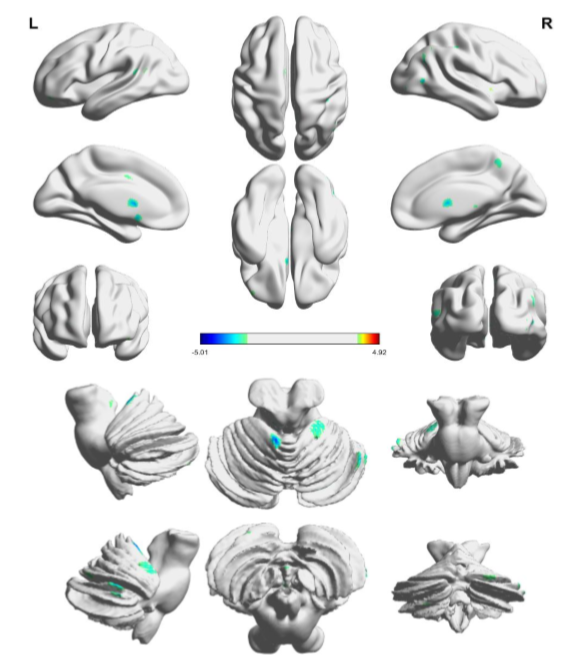
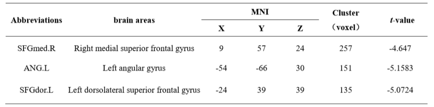
TABLE 2 | Brain areas with decreased functional connectivity in the AUD group compared with the HC group. Abbreviations: AUD, alcohol use disorder; HC, healthy controls; MNI, montreal neurological institute.