2696
Brain regional connectome-wide search identified a resting-state functional connectivity locus within precunes associated with rumination symptom severity in mood and anxiety disorders1Laureate Institute for Brain Research, Tulsa, OK, United States, 2Japan Society for the Promotion of Science, Tokyo, Japan, 3Department of Electrical and Computer Engineering, University of Oklahoma, Tulsa, OK, United States, 4Stephenson School of Biomedical Engineering, University of Oklahoma, Norman, OK, United States
Synopsis
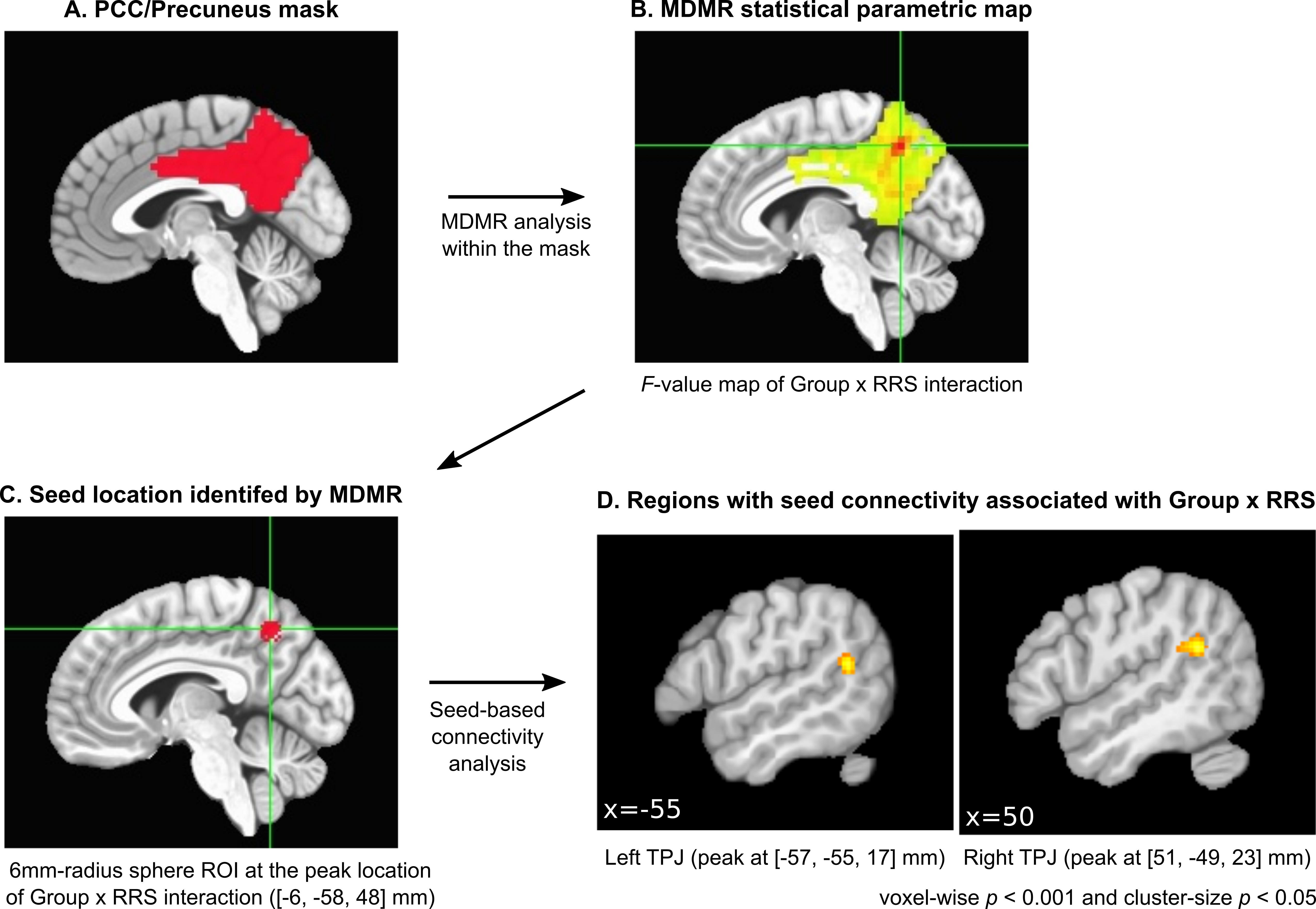
We identified a precise locus within the precuneus that has resting-state functional connectivity (rsFC) associated with rumination symptom severity for mood and anxiety (MA) disorder patients. We devised brain regional connectome-wide association analysis, which used multivariate distance matrix regression for searching voxels with connectivity correlated with the Ruminative Responses Scale (RRS) within the posterior cingulate cortex and the precuneus. The analysis identified voxels in the precuneus having rsFC significantly associated with RRS. Functional connectivity between the precuneus and bilateral temporoparietal junction (TPJ) had a significant positive correlation with RRS in MA patients but not in the healthy participants.
Purpose
Rumination is a common and debilitating symptom of mood and anxiety disorders1. The posterior cingulate cortex (PCC) and precuneus - as parts of the default mode network (DMN) - have been implicated in ruminative thought, as the DMN is associated with internal self-oriented thought and PPC/precuneus hyperactivity in resting-state is often reported for patients with mood disorders2. However, the PCC and precuneus, as well as the DMN, are not homogeneous regions3,4 and which specific parts of the PCC/precuneus and DMN are responsible for pathological rumination remains to be determined. The present study performed an exploratory analysis to identify a locus in the PCC/precuneus that has resting-state functional connectivity (rsFC) associated with rumination symptom severity for mood and/or anxiety disorder patients.Methods
The study was performed on samples from the Tulsa 1000 study5. Data included 46 healthy control (HC: 24 females, age 18-52) and 225 mood and/or anxiety disorder (MA: 188 females, age 18-55) individuals. Resting-state fMRI data (TR/TE=2000/27ms, 8min) was processed with AFNI (https://afni.nimh.nih.gov/); discarding initial 5 volumes, despike, RETROICOR, slice-timing and motion corrections, spatial normalization to MNI template with ANTs6, spatial smoothing with FWHM=6mm kernel, and scaling signal to percent change. GLM analysis regressed out the effects of six motion parameters, their temporal derivatives, three principal components of ventricle signal, local white matter average signal (ANATICOR7), and slow fluctuations modeled by Legendre polynomials. The residual signal of the GLM analysis was a subject to the following analysis.
We introduced a brain regional connectome-wide approach to search the region within the PCC/precuneus having rsFC associated with the Ruminative Responses Scale (RRS)8. A whole-brain connectivity analysis using multivariate distance matrix regression (MDMR)9,10 was performed for the seed voxels within a target area. Notably, limiting the search area could improve the sensitivity of the analysis due to fewer multiple comparisons, as well as vastly reduce computational cost compared to whole-brain connectome-wide analysis. Figure 1 shows the analysis procedure. We made a mask of PCC and precuneus areas using the Desikan-Killiany atlas11 (Fig. 1A). MDMR analysis was done for the voxels within the mask. In MDMR, correlation of signal time-courses for a seed voxel to all other brain voxels was calculated and applied the Fisher z-transformation to make a connectivity map. A distance matrix between connectivity maps of subjects was applied nonparametric MANCOVA with a design matrix including regressors of group (HC, MA), RRS score, their interaction, age, gender, and motion. P-value was evaluated by a 10,000-repetition permutation test. These steps were repeated for each voxel as a seed to make a statistical parametric map. MDMR statistical parametric map (Fig. 1B) indicates an association between a regressor and a multivariate whole-brain connectivity pattern at a voxel. Since the MDMR result does not show which specific connectivity was associated with a regressor, post-hoc seed-based connectivity analysis was performed for the significant region of the MDMR statistical map (Fig. 1C). We used univariate voxel-wise GLM analysis for rsFC at the seed with the same design matrix as MDMR for the post-hoc analysis.
Results
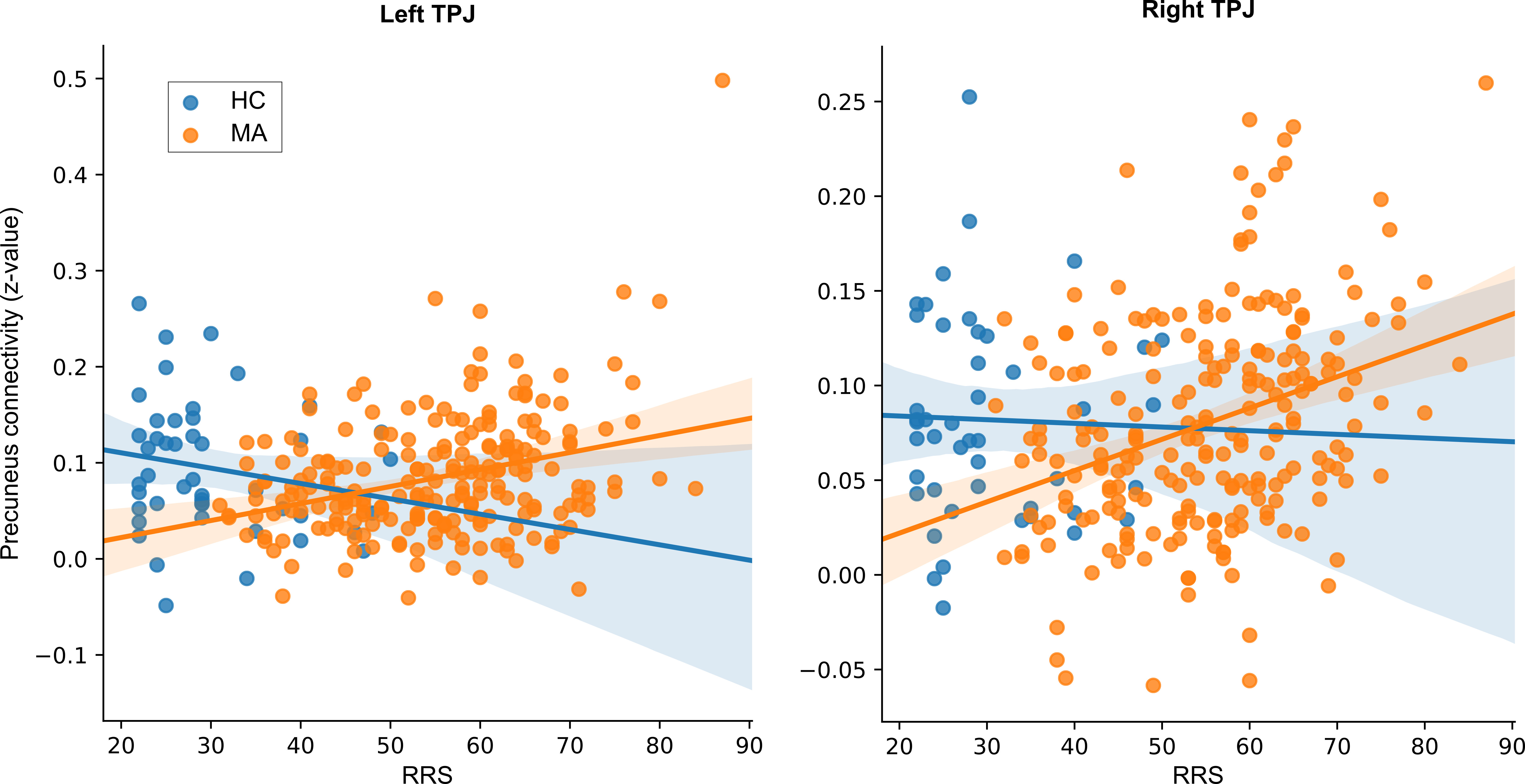
MDMR analysis found a significant interaction effect of group×RRS (p<0.005) in the precuneus (Fig. 1B). No other significant effect was found. Post-hoc analysis was performed for the locus (seed ROI, 6mm-radius sphere) of the MDMR statistical parametric map (Fig. 1C). The analysis revealed that rsFC between the precuneus and the bilateral temporoparietal junction (TPJ) regions had a significant interaction effect (p<0.001 and cluster-size p<0.05). Fig. 2 shows that connectivity between the precuneus and TPJ had significant positive correlation with RRS for MA group (left; t[263]=4.82, p<0.001, right; t[263]=4.94, p<0.001) but not for HC group (left; t[263]=-1.56, p=0.120, right; t[263]=-0.43, p=0.669).Discussion
Brain regional connectome-wide analysis identified a precise location within the precuneus that has resting-state functional connectivity associated with rumination severity in MA individuals. The identified locus in the precuneus had increased connectivity with TPJ and correlated with the severity of rumination symptoms. TPJ is a part of DMN and has been implicated in social cognitive functions, including theory of mind, sense of agency, and self-other differentiation12. The hyperconnectivity between the precuneus and the TPJ associated with rumination severity suggests that altered self-referential processing might underpin pathological rumination.
This localization is not only crucial for functional mapping but also useful for identifying locus of targeted intervention, such as real-time fMRI neurofeedback or transcranial magnetic stimulation. The introduced approach can have wide and important applications for target discovery of novel intervention in psychiatric disorders.
Acknowledgements
This work was supported by Laureate Institute for Brain Research, the William K. Warren Foundation, and National Institute of General Medical Sciences, National Institutes of Health Award 1P20GM121312.References
1. Nolen-Hoeksema, S., The role of rumination in depressive disorders and mixed anxiety/depressive symptoms. Journal of abnormal psychology, 2000;109(3):504.
2. Hamilton, J.P., M. Farmer, P. Fogelman, et al., Depressive Rumination, the Default-Mode Network, and the Dark Matter of Clinical Neuroscience. Biol Psychiatry, 2015;78(4):224-30.
3. Leech, R., R. Braga, and D.J. Sharp, Echoes of the brain within the posterior cingulate cortex. J Neurosci, 2012;32(1):215-22.
4. Leech, R., S. Kamourieh, C.F. Beckmann, et al., Fractionating the default mode network: distinct contributions of the ventral and dorsal posterior cingulate cortex to cognitive control. J Neurosci, 2011;31(9):3217-24.
5. Victor, T.A., S.S. Khalsa, W.K. Simmons, et al., Tulsa 1000: a naturalistic study protocol for multilevel assessment and outcome prediction in a large psychiatric sample. BMJ Open, 2018;8(1):e016620.
6. Avants, B.B., C.L. Epstein, M. Grossman, et al., Symmetric diffeomorphic image registration with cross-correlation: evaluating automated labeling of elderly and neurodegenerative brain. Med. Image Anal, 2008;12(1):26-41.
7. Jo, H.J., Z.S. Saad, W.K. Simmons, et al., Mapping sources of correlation in resting state FMRI, with artifact detection and removal. NeuroImage, 2010;52(2):571-82.
8. Treynor, W., R. Gonzalez, and S. Nolen-Hoeksema, Rumination reconsidered: A psychometric analysis. Cognitive therapy and research, 2003;27(3):247-259.
9. Shehzad, Z., C. Kelly, P.T. Reiss, et al., A multivariate distance-based analytic framework for connectome-wide association studies. Neuroimage, 2014;93 Pt 1:74-94.
10. Misaki, M., R. Phillips, V. Zotev, et al., Connectome-wide investigation of altered resting-state functional connectivity in war veterans with and without posttraumatic stress disorder. Neuroimage Clin, 2018;17:285-296.
11. Desikan, R.S., F. Ségonne, B. Fischl, et al., An automated labeling system for subdividing the human cerebral cortex on MRI scans into gyral based regions of interest. Neuroimage, 2006;31(3):968-980.
12. Eddy, C.M., The junction between self and other? Temporo-parietal dysfunction in neuropsychiatry. Neuropsychologia, 2016;89:465-477.
Figures