2692
Altered functional connectivity and spectroscopic metabolites related to treatment response in adolescents with bipolar disorder1Radiology, West China Hospital of Sichuan University, Chengdu, China
Synopsis
The reason for the inconsistency of bipolar disorder (BD) patients’ brain functional status and metabolic levels of treatment response is still not clear. This task-based fMRI study was carried out to figure out the relationship between medication treatment and brain status in function and metabolites. By analyzing functional connectivity and correlating metabolic markers in treatment response and no response BD patients, we found medication can affect the brain functional status and metabolic level in BD patients, and precentral gyrus is a key region during BD illness course.
Introduction
The heterogeneity of bipolar disorder (BD) patients has been confirmed by previous studies, and inconsistent results in patient’s response to medication also exist. 1-2 However, it is still not clear about the reasons of inconsistency. The question of specific differences between the brain function and metabolic status of patients respond and not respond to medication remains to be solved. At the same time, the relationship between metabolic makers and imaging functional makers is also confused. Therefore, in order to understand the difference of brain functional status and metabolic level between treatment responders and non-responders. A task-based fMRI study with 49 firs-episode BD adolescents was carried to figure out the questions above.Methods
Forty-nine first-episode adolescents with BD (mean age: 14.09±1.81 years) and fifty-eight matched healthy controls (mean age:14.59±2.98 years) were recruited. Continuous Performance Task (CPT) was applied during fMRI scans on a 4-T Varian Unity INOVA scanner at baseline and after 6 weeks’ antipsychotic treatment with quetiapine. Scores of Young Mania Rating Scale (YMRS) at baseline and endpoint were collected, and the treatment response was defined by the Ratio = (YMRStreated-YMRSbaseline) / YMRStreated≥0.5. Anterior cingulate cortex (ACC), left ventrolateral prefrontal cortex (LVLPFC) and right ventrolateral prefrontal cortex (RVLPFC) were selected as 3 seeds to acquire metabolic data by magnetic resonance spectroscopy (MRS). Functional connectivity (FC) between regions-of-interest (ROI) and the whole brain voxels were calculated by using DPASF software (Data Processing Assistant for Resting-State fMRI, version 2.3; http://rfmri.org/DPARSF) and REST software (Resting-State fMRI Data Analysis Toolkit V1.8; http://restfmri.net/forum/rest). 3 Statistical analyses were performed with SPSS Statistics 22.0 software.Results
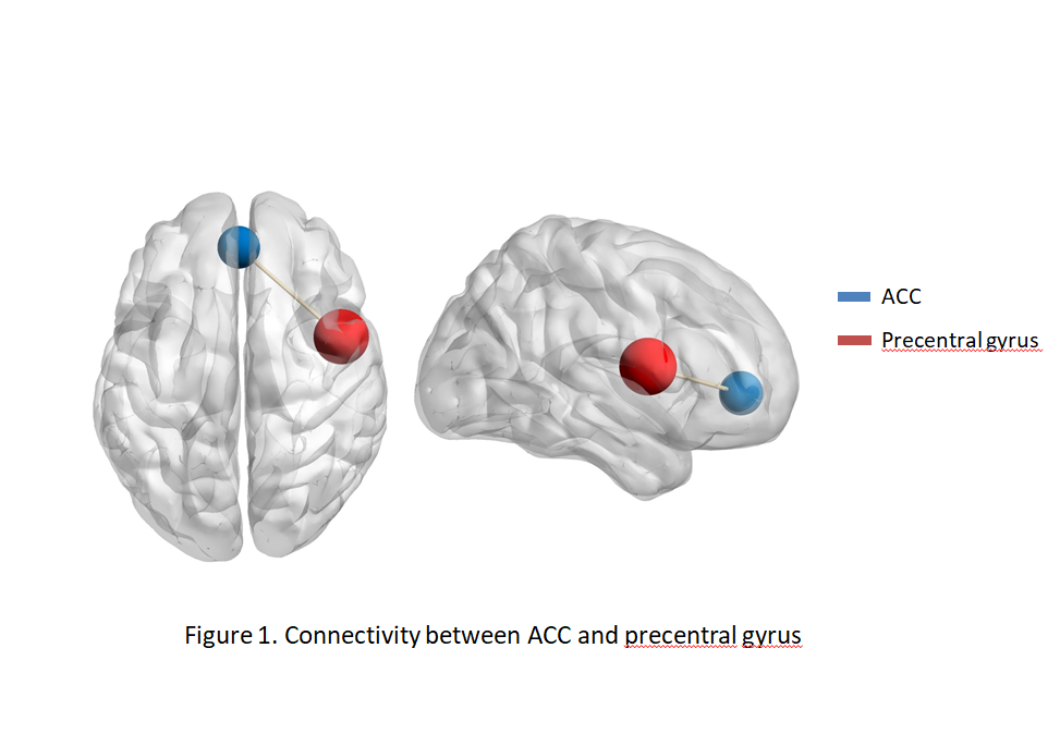
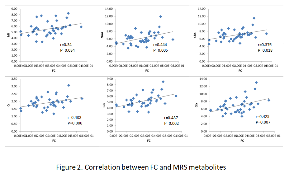
No significant FC difference exhibited in patients compared with healthy controls before treatment. After 6 weeks’ antipsychotic treatment with quetiapine, 79.59% patients (39 patients) respond to the medication, and 10 patients showed no response to treatment. The Analysis of Variance (ANOVA) test of FC based on seed were performed among responders, non-responders and healthy control groups. We found significant FC decrease between ACC and right precentral gyrus both in responders and non-responders compared with healthy controls, and responders showed higher FC compared with non-responders (corrected by GRF, P<0.01) (Figure 1). In responder group, positive correlation were detected between FC and MRS metabolites, including myo-inositol (MI), N-acetyl aspartate (NAA), choline (Cho), creatine (Cr) Glutamate (Glu) and glutamate+glutamine (Glx) (Figure 2). There is no significant correlation between FC and MRS metabolites in non-responder group.Discussion
After 6 weeks of antipsychotic treatment with quetiapine, most of patients improved in clinical assessment. Both responder and non-responder groups showed lower FC than healthy controls between ACC and right precentral gyrus. Previous studies have demonstrated extensive functional dysconnectivity and grey matter reduction in BD patients both in youths and adults, and the precentral gyrus related circuits are among the brain areas with functional changes. 4-5 Our FC findings showed the alternation in the same brain region. The FC change of precentral gyrus could link to attentional processing, and the stimulated alternation under CPT task amplify the disruption occurs in ACC-precentral gyrus circuits. The decreased imaging maker is continuous with FC findings in meta-analyses with BD and schizophrenia.6 FC changes involved in precentral gyrus may be a phenotype for mental illness. Higher FC in responder group was detected. This alternation could be related to the effects of quetiapine treatment.
Antipsychotic treatment may work via modulating the neurometabolites in youth with BD.7 Those metabolites are important markers reflecting metabolic environment in cerebrum. NAA is a marker for neuronal integrity and is exclusively found in the brain. Glu is the primary excitatory neurotransmitter in the central nervous. The alternation of NAA, Glu and other makers have been demonstrated in mental illness,8 the behavior of congruent change of metabolites above reflected this alternation could be accompanied with treatment effects.
Conclusion
Treatment of quetiapine can affect the brain functional status and metabolic level in BD patients, and precentral gyrus is a key region which can reflect the brain changes in multi-aspect during BD illness course.Acknowledgements
No acknowledgement found.References
1. Hibar DP, Westlye LT, Doan NT, Jahanshad N, Cheung JW, Ching CRK, Versace A, Bilderbeck AC, Uhlmann A, Mwangi B, Krämer B, Overs B, Hartberg CB, Abé C14, Dima D, Grotegerd D, Sprooten E ,Bøen E, Jimenez E, Howells FM, Delvecchio G, et al. Cortical abnormalities in bipolar disorder: an MRI analysis of 6503 individuals from the ENIGMA Bipolar Disorder Working Group. Mol Psychiatry. 2018 Apr;23(4):932-942.
2. Lencer R, Yao L, Reilly JL, Keedy SK, McDowell JE, Keshavan MS, Pearlson GD, Tamminga CA, Gershon ES, Clementz BA, Lui S, Sweeney JA. Alterations in intrinsic fronto-thalamo-parietal connectivity are associated with cognitive control deficits in psychotic disorders. Hum Brain Mapp. 2018 Sep 10. doi: 10.1002/hbm.24362. [Epub ahead of print]
3. Zang YF, He Y, Zhu CZ, Cao QJ, Sui MQ, Liang M, Tian LX, Jiang TZ, Wang YF. Altered baseline brain activity in children with ADHD revealed by resting-state functional MRI. Brain Dev. 2007 Mar;29(2):83-91.
4. Zhang W, Xiao Y, Sun H, Patino LR, Tallman MJ, Weber WA, Adler CM, Klein C, Strawn JR, Nery FG, Gong Q, Sweeney JA, Lui S, DelBello MP. Discrete patterns of cortical thickness in youth with bipolar disorder differentially predict treatment response to quetiapine but not lithium. Neuropsychopharmacology. 2018 Oct;43(11):2256-2263.
5. Wolfers T, Doan NT, Kaufmann T, Alnæs D, Moberget T, Agartz I, Buitelaar JK, Ueland T, Melle I, Franke B, Andreassen OA, Beckmann CF, Westlye LT, Marquand AF. Mapping the Heterogeneous Phenotype of Schizophrenia and Bipolar Disorder Using Normative Models. JAMA Psychiatry. 2018 Oct 10. doi: 10.1001/jamapsychiatry.2018.2467. [Epub ahead of print]
6. Kraguljac NV, Reid M, White D, Jones R, den Hollander J, Lowman D, Lahti AC. Neurometabolites in schizophrenia and bipolar disorder - a systematic review and meta-analysis. Psychiatry Res. 2012 Aug-Sep;203(2-3):111-25.
7. Strawn JR, Patel NC, Chu WJ, Lee JH, Adler CM, Kim MJ, Bryan HS, Alfieri DC, Welge JA, Blom TJ, Nandagopal JJ, Strakowski SM, DelBello MP. Glutamatergic effects of divalproex in adolescents with mania: a proton magnetic resonance spectroscopy study. J Am Acad Child Adolesc Psychiatry. 2012 Jun;51(6):642-51.
8. Soeiro-de-Souza MG, Otaduy MCG, Machado-Vieira R, Moreno RA, Nery FG, Leite C, Lafer B. Anterior Cingulate Cortex Glutamatergic Metabolites and Mood Stabilizers in Euthymic Bipolar I Disorder Patients: A Proton Magnetic Resonance Spectroscopy Study. Biol Psychiatry Cogn Neurosci Neuroimaging. 2018 Mar 31. pii: S2451-9022(18)30076-4. doi: 10.1016/j.bpsc.2018.02.007. [Epub ahead of print]
Figures