2690
Altered white matter microstructure correlates with cognitive functions in children and adolescents with bipolar disorder1Department of Electrical and Systems Engineering, School of Engineering and Applied Science, University of Pennsylvania, Philadelphia, PA, United States, 2Department of Radiology, Children’s Hospital of Philadelphia, Philadelphia, PA, United States, 3Department of Bioengineering, School of Engineering and Applied Science, University of Pennsylvania, Philadelphia, PA, United States, 4Baylor College of Medicine-Texas Children’s Hospital, Houston, TX, United States, 5Department of Radiology, Perelman School of Medicine, University of Pennsylvania, Philadelphia, PA, United States
Synopsis
Cognitive impairments and white matter (WM) microstructural alterations have been found in subjects with bipolar disorder (BD). However, the relationship between WM microstructural alterations and impulsivity, a prominent cognitive trait, in children/adolescents with BD is not known. In this study, diffusion MRI and cognitive assessments were obtained from 19 children/adolescents diagnosed with BD and 23 age-matched healthy controls. We found increased radial diffusivity(RD), reflecting disrupted myelin, in major WM tracts such as corpus callosum. Significant correlation between RD in WM tracts regulating impulsivity and response time to affective words was found, suggesting the association between WM myelin disruption and impulsivity.
Purpose
Cognitive impairments and white matter (WM) microstructural alterations have been found in subjects with bipolar disorder (BD)[1-3]. However, the relationship between WM microstructural alterations and impulsivity, a prominent cognitive trait, in children and adolescents with BD is not known. In this study, diffusion MRI and cognitive assessments were obtained from 19 children and adolescents diagnosed with BD and 23 age-matched healthy controls. We aimed to identify the WM tracts with increased radial diffusivity (RD) which reflects disrupted myelin and aimed to delineate the correlation between increased RD in WM tracts and impairment of cognitive functions in children and adolescents with BD.Methods
Subjects and data acquisition: 19 children and adolescents diagnosed with BD (aged 12.1 ± 3.5 years, Male/Female = 7/12) and 23 age-matched healthy controls (HC) (aged 12.0 ± 3.4 years, Male/Female = 11/12) were recruited. Children and adolescents were diagnosed with type I, type II, or not otherwise specified (NOS) bipolar disorder based on the Mini International Neuropsychiatric Interview-KID (MINI) and the Course and Outcome of Bipolar Youth (COBY) Criteria. All MRI images were acquired on a 3T Philips Ingenia scanner. Diffusion weighted images (DWI) were acquired with the following parameters: TR/TE = 12400/77 ms; non-zero b-value of 1000 s/mm2 with 21 independent diffusion gradient directions; FOV = 256x256 mm2; in-plane imaging resolution=2x2 mm2; slice thickness = 3 mm without slice gap; slice number = 44. DWI data of 16 BD and 22 HC was used for the following analysis after discarding DWI of three BD subjects and one HC subject due to incomplete acquisition or severe motion artifacts. All subjects went through Cambridge Neuropsychological Test Automated Battery (CANTAB) cognition tests to assess their cognitive functions[4]. In Affective Go/No-Go (AGN) task, subjects were asked to select a word when it matched the affective categories (positive or negative), and their response latencies in correct trials were recorded. One BD and four HC subjects’ latencies to both positive and negative stimuli, and one HC subject’s latency to negative stimuli in AGN tasks were not successfully recorded. Measurements of DTI-derived metrics on WM skeleton: Head motion and eddy-current correction were performed by registering all DWI images to b0 image using affine transformation in DTIstudio (http://www.MRIstudio.org). Diffusion tensor was then fitted and four DTI-derived metrics, fractional anisotropy (FA), RD, axial diffusivity (AD), and mean diffusivity (MD) were calculated in DTIstudio. To reduce the partial volume effect, Tract-Based Spatial Statistic (TBSS) from FSL was performed (https://fsl.fmrib.ox.ac.uk/fsl/fslwiki/TBSS). Specifically, the core of WM tracts, namely WM skeleton, was extracted from the averaged FA maps after registration to the JHU ICBM FA template. Skeletonized FA, RD, AD, and MD maps were thus obtained. Comparison between BD and HC and correlation with cognitive assessments: Voxel-wise comparison between BD and HC was carried out with skeletonized DTI-derived maps using permutation-based nonparametric statistics (Randomise function in FSL) with 5000 permutations. The threshold free cluster enhancement (TFCE in FSL) at cluster level threshold of p < 0.05 was applied. Age effect was excluded by setting age as a covariate in comparison. A digital WM atlas JHU ICBM-DTI-81 (http://cmrm.med.jhmi.edu) was used to label the voxels with significant difference to corresponding WM tracts. Skeletonized DTI-derived metrics in voxels with significant difference in each tract were linearly fitted to cognitive assessments from CANTAB.Results
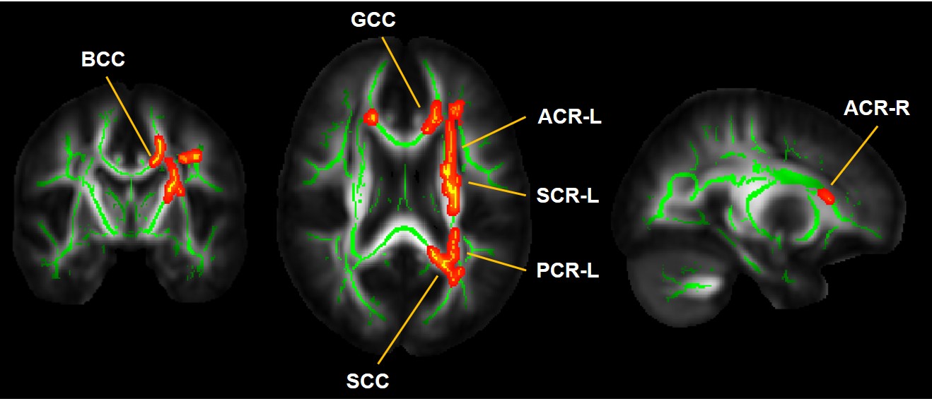
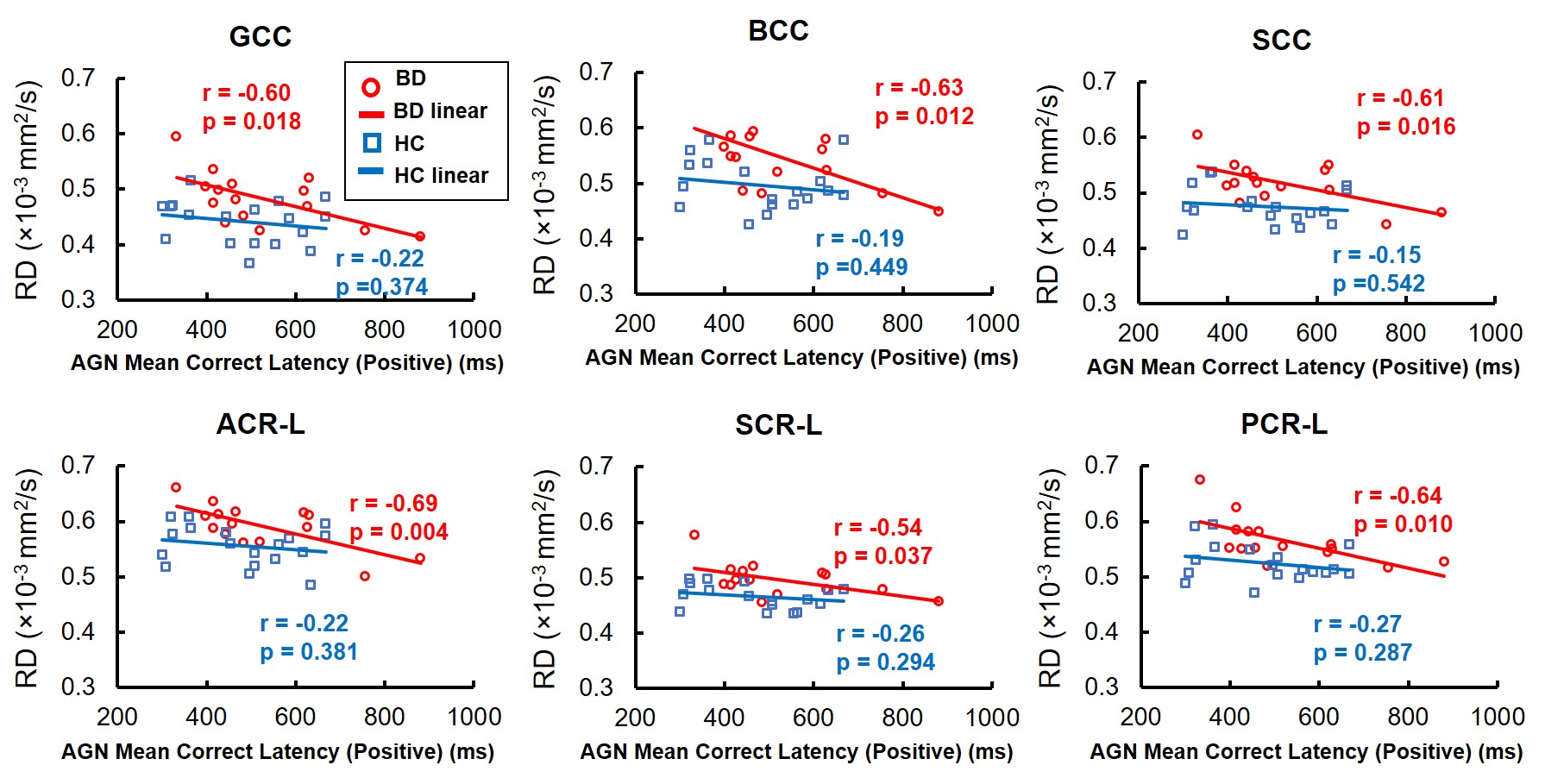
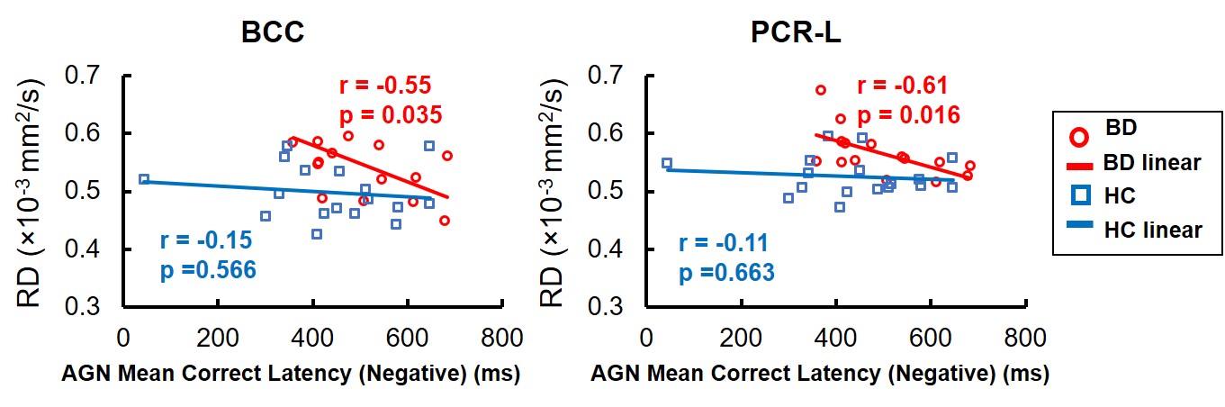
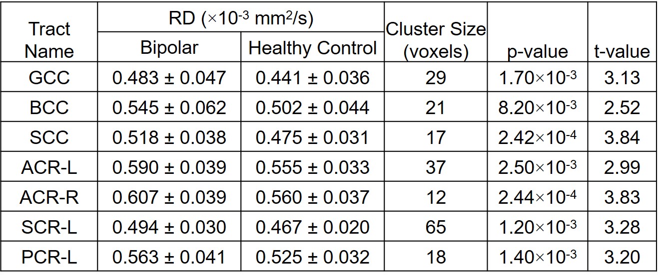
We found significantly elevated RD (p<0.05) in children and adolescents with BD in clusters in the corpus callosum (CC), and left and right corona radiata (CR-L and CR-R) (Figure 1 and Table 1). We did not find significant difference in other DTI-derived metrics. More importantly, RD values in identified clusters from CC and CR-L significantly correlated with response time to positive and negative stimuli in the AGN tasks (p<0.05) (Figure 2 and 3). The shorter the response time was, the larger the RD was. In HC, this correlation was not significant.Discussion and Conclusion
Higher RD compared with HC were found distributed mainly in the CC and CR in the children and adolescents with BD, consistent with previous studies[3,5]. Increased RD suggests disrupted myelin in these WM tracts in the children and adolescents with BD[6]. Furthermore, abnormally higher RD in GCC and ACR-L which regulate impulsivity[7] significantly correlated with reduced response time to affective words in Affective Go/No-Go tasks, suggesting the association between WM myelin disruption and increased impulsivity. The significant correlations between response time and DTI metrics provide preliminary evidence of microstructural underpinning of the impairment of cognitive functions for children and adolescents with BD.Acknowledgements
This study is funded by NIH MH092535, MH092535-S1 and HD086984.References
[1] Bauer, I. E., Frazier, T. W., Meyer, T. D., Youngstrom, E., Zunta–Soares, G. B., & Soares, J. C. (2015). Affective processing in pediatric bipolar disorder and offspring of bipolar parents. Journal of child and adolescent psychopharmacology, 25(9), 684-690.
[2] Lewandowski, K. E., Ongür, D., Sperry, S. H., Cohen, B. M., Sehovic, S., Goldbach, J. R., & Du, F. (2015). Myelin vs axon abnormalities in white matter in bipolar disorder. Neuropsychopharmacology, 40(5), 1243.
[3] Barysheva, M., Jahanshad, N., Foland-Ross, L., Altshuler, L. L., & Thompson, P. M. (2013). White matter microstructural abnormalities in bipolar disorder: a whole brain diffusion tensor imaging study. NeuroImage: clinical, 2, 558-568.
[4] Wu, M. J., Passos, I. C., Bauer, I. E., Lavagnino, L., Cao, B., Zunta-Soares, G. B., & Soares, J. C. (2016). Individualized identification of euthymic bipolar disorder using the Cambridge Neuropsychological Test Automated Battery (CANTAB) and machine learning. Journal of affective disorders, 192, 219-225.
[5] Bollettini, I., Poletti, S., Locatelli, C., Vai, B., Smeraldi, E., Colombo, C., & Benedetti, F. (2015). Disruption of white matter integrity marks poor antidepressant response in bipolar disorder. Journal of affective disorders, 174, 233-240.
[6] Song, S. K., Sun, S. W., Ramsbottom, M. J., Chang, C., Russell, J., & Cross, A. H. (2002). Dysmyelination revealed through MRI as increased radial (but unchanged axial) diffusion of water. Neuroimage, 17(3), 1429-1436.
[7] McDonald, V., Hauner, K. K., Chau, A., Krueger, F., & Grafman, J. (2017). Networks underlying trait impulsivity: Evidence from voxel‐based lesion‐symptom mapping. Human brain mapping, 38(2), 656-665.
Figures