2687
The Importance of Identifying Functional Val158Met Polymorphism in Catechol-O- Methyltransferase (COMT) when Assessing MRI-based Volumetric Measurements in Major Depressive Disorder1Biomedical Engineering, Stony Brook University, Stony Brook, NY, United States, 2Psychiatry, Stony Brook Medicine, Stony Brook, NY, United States, 3Radiology, Stony Brook Radiology, Stony Brook, NY, United States
Synopsis
Using voxel-based morphology we investigated the relationship between COMT gene polymorphism and volumetric abnormalities in major depressive disorder patients and healthy controls. A significant difference in the right hippocampus (p=0.015) was found between the interaction of diagnosis and genotype, which suggests that COMT polymorphism must be considered during any volumetric analysis for depression.
Introduction
Major depressive disorder (MDD) affects 350 million people worldwide and is a debilitating chronic disease that impacts 1 in 6 people in the United States during their1,2. Depression is a disorder based on heterogeneous symptomology which makes it harder to detect and cure3. The hippocampal region has been found to be associated with MDD, with patients showing volumetric reductions in hippocampal size compared to healthy controls (HC)4-8.
The COMTval158met polymorphism produces a single nucleotide polymorphism (SNP)-rs4680- at codon 158, from G to A, resulting in a methionine (Met) amino acid instead of a valine (Val) amino acid. Previous studies on healthy controls have found that Val/Val homozygous COMT genotype has been associated with decreased hippocampal and amygdala volumes in brain scans9. Therefore, we hypothesize that the COMT Val allele individuals with MDD will have the greatest volumetric reductions in the hippocampus and amygdala compared to COMT Met allele subjects.
Methods
Data used in this project were anonymized and previously acquired in an IRB approved study. 60 subjects met the DSM-IV criteria for MDD (37 females, 23 males) and 25 healthy controls (12 females, 13 males) were included in this analysis. The acquisition of magnetic resonance imaging (MRI) was performed as previously stated10. Volumetric analysis was performed using Freesurfer 5.3. The hippocampus and amygdala were chosen because of prior referencing of their role in depression and their volumetric differences in healthy Val/Val subjects. DNA was extracted from lymphocytes and epithelial cells of the cheek using either a Puregene Kit or BuccalAmp DNA Extraction Kit11. The polymerase chain reaction-restriction fragment length (PCR-RFLP) was used to identify the COMT genotype. Although MDD patients and controls differed in terms of age, both linear regression and matched samples were used. The data analysis was performed on SPSS for Windows, Version 23 (SPSS Inc., Chicago, IL, USA).Results
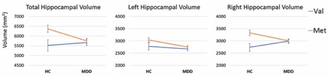
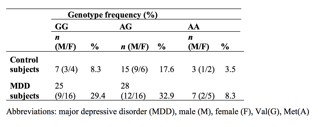
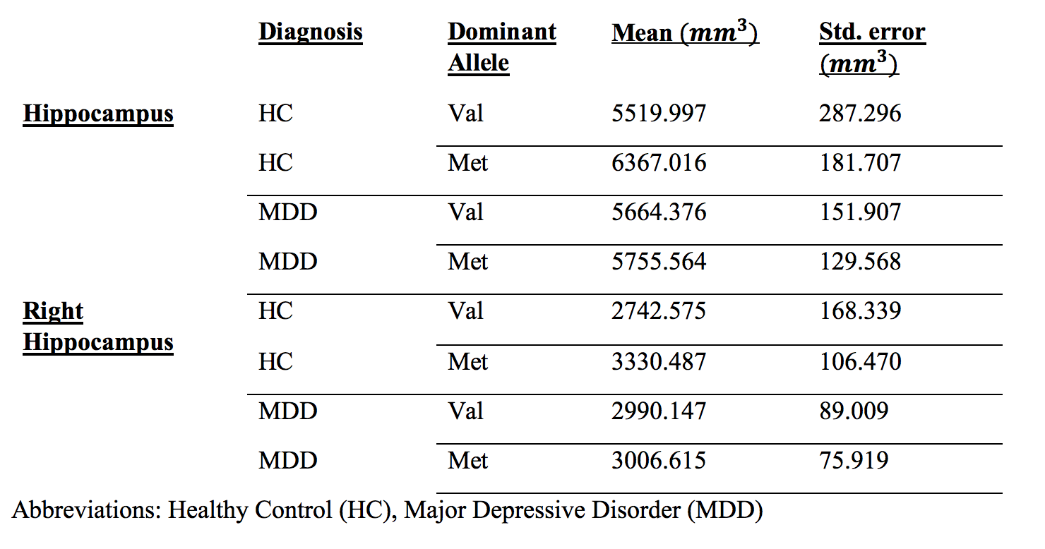
Due to the intrinsic limitations of the data set, there is an age difference between MDD patient and healthy control groups, (Healthy Control=33.6, MDD = 40.3, p-value= 0.031). There were three genotypes: homozygous Val (GG; n= 32), homozygous Met (AA; n=10) and heterozygous Met/Val (AG; n=43). No significant differences in genotype frequencies were identified between the MDD patients and the healthy control group (χ2=0.06, df=2, p=0.970). Of the three genotypes, they were subsequently paired as Met allele (Met/Met & Val/Met) (A; n=53) and Val allele (Val/Val) (G; n =32). Between the 25 HCs and 60 MDD subjects we found no significant difference in volumes between MDD and HC. We found a significant difference between the interaction of diagnosis and genotype in the right hippocampal volume (p=0.015). No significant difference was found for either the amygdala (p=0.558) or the entire hippocampus (p=0.059). Figure 1 shows the mean volume of the left, right and entire hippocampus between subject diagnosis and combined genotype. Table 2 below shows that the mean volume of the right hippocampus of HC.Discussion
In our Met dominant model, our data showed that HC Val genotype subjects had lower hippocampal volumes compared to HC Met genotype, which agrees with previous studies9. We did not found significant difference in means between the interaction of diagnosis and paired genotypes in either the amygdala (p=0.558) or the hippocampus (p=0.059). Although not significant, Val genotype subjects with MDD, whom we hypothesized to have the lowest mean hippocampal volumes across all groups, had the highest hippocampal volume. Although no significance was found in entire hippocampal volume, we did find significance in right hippocampal volume. We can clearly see in Figure 1a-b that the right hippocampal volumes of MDD patients differ depending on the certain polymorphism that they have; where HC Val dominant genotypes have smaller right hippocampal volumes compared to MDD counterparts, but HC Met dominant genotypes have larger right hippocampal volumes than their MDD counterparts. While some studies have found a statistically significant volumetric difference within the hippocampus of depressed patients12, there are others that see no difference in hippocampal volumes in adults13. Our study suggest that it is important for researchers to take COMT polymorphism into account when measuring volumetric differences in hippocampal volumes since our data suggests that results may vary due to genetic variables such as COMT polymorphism.Conclusion
This study has provided insight to a possible association between the COMT val158met polymorphism and volumetric difference in major depressive disorder patients. Our data suggests that there is a significant difference between the interaction of the COMT val158met polymorphism and diagnosis when studying hippocampal volumes in MDD.Acknowledgements
No acknowledgement found.References
1. R. C. Kessler, P. Berglund, O. Demler, R. Jin, K. R. Merikangas and E. E. Walters, Archives of general psychiatry 62 (6), 593-602 (2005).
2. W. H. Organization, World Mental Health Day 10 (2012).
3. M. ten Have, F. Lamers, K. Wardenaar, A. Beekman, P. de Jonge, S. van Dorsselaer, M. Tuithof, M. Kleinjan and R. de Graaf, Journal of affective disorders 190, 395-406 (2016).
4. F. P. MacMaster and V. Kusumakar, BMC medicine 2 (1), 2 (2004).
5. Y. I. Sheline, Biological psychiatry 48 (8), 791-800 (2000).
6. J. Cole, A. W. Toga, C. Hojatkashani, P. Thompson, S. G. Costafreda, A. J. Cleare, S. C. Williams, E. T. Bullmore, J. L. Scott and M. T. Mitterschiffthaler, Journal of affective disorders 126 (1-2), 272-277 (2010).
7. C. Eker and A. S. Gonul, (Taylor & Francis, 2010).
8. G. M. MacQueen, K. Yucel, V. H. Taylor, K. Macdonald and R. Joffe, Biological psychiatry 64 (10), 880-883 (2008).
9. W. D. Taylor, S. Züchner, M. E. Payne, D. F. Messer, T. J. Doty, J. R. MacFall, J. L. Beyer and K. R. R. Krishnan, Psychiatry Research: Neuroimaging 155 (2), 173-177 (2007).
10. R. V. Parsey, R. T. Ogden, J. M. Miller, A. Tin, N. Hesselgrave, E. Goldstein, A. Mikhno, M. Milak, F. Zanderigo and G. M. Sullivan, Biological psychiatry 68 (2), 170-178 (2010).
11. H. A. Erlich, PCR technology. (Springer, 1989).
12. J. D. Bremner, M. Narayan, E. R. Anderson, L. H. Staib, H. L. Miller and D. S. Charney, American Journal of Psychiatry 157 (1), 115-118 (2000).
13. R. S. Hastings, R. V. Parsey, M. A. Oquendo, V. Arango and J. J. Mann, Neuropsychopharmacology 29 (5), 952 (2004).
Figures