2686
Hippocampus-related regional and network functional deficits in first-episode drug-naïve major depressive disorder: a resting-state functional MRI study1Huaxi MR Research Center (HMRRC), Department of Radiology, West China Hospital of Sichuan University, Chengdu, China, Chengdu, China
Synopsis
Previous neuroimaging studies have suggested that major depressive disorder (MDD) may be correlated with changes in regional- or network-level brain function. The purposes of the present study were to investigate changes of amplitude of low-frequency fluctuation (ALFF) and functional connectivity (FC) in bilateral hippocampus by resting-state functional magnetic resonance imaging (rs-fMRI) in first-episode drug-naive major depressive disorder (MDD) patients. Our findings demonstrate that the hippocampus and dACC contribute to the underlying pathophysiology of MDD at an early-stage.
Purpose
Previous neuroimaging studies have suggested that major depressive disorder (MDD) may be correlated with changes in regional- or network-level brain function1-2. However, few studies have explored the resting-state cerebral functional alternations in first-episode drug-naïve MDD patients at both the regional and network levels, particularly focusing on the hippocampus. Herein, we aimed to investigate changes of amplitude of low-frequency fluctuation (ALFF)3, as a measure of regional brain activity, and functional connectivity (FC) in bilateral hippocampus by resting-state functional magnetic resonance imaging (rs-fMRI) in first-episode drug-naive MDD patients, then to determine whether these alterations are correlated with the clinical measures of MDD.Methods
The study was approved by the local ethical committee and written informed consent was obtained from all subjects. A total of 30 first-episode drug-naive MDD patients who met DSM-IV criteria for MDD and 52 age, gender and handedness well matched healthy controls were recruited. Depressive symptom severity was assessed using the 17-item Hamilton Depression Rating Scale (HAMD). The rs-fMRI sensitized to changes in the blood oxygen level dependent (BOLD) signal (repetition time/echo time = 2,000/30 msec; flip angle=90 degrees) were obtained with a gradient-echo echo-planar imaging (EPI) sequence via a 3.0 T GE scanner. Participants were instructed to relax with their eyes closed and keep still. The ALFF maps were calculated using DPARSF software (http://www.restfmri.net) for each subject. The region of interests (ROIs) seed-based approach was used to assess the resting-state FC of the right hippocampus and left hippocampus. The ROIs of the right and left hippocampus were defined according to the automated anatomical labeling (AAL) template4 contained in REST (http://resting-fmri.sourceforge.net/). The correlation analyses were performed voxel-wise between each seed and the rest of the brain. Finally, the correlation coefficients in each voxel were transformed to z values using the Fisher r-to-z transformation to improve normality. Afterward, voxel-based and seed-to-voxel analyses of the ALFF and FC differences between the MDD and HCS were performed separately using the two-sample t-test in SPM8 (http://www.fil.ion.ucl.ac.uk/spm). The significance threshold was set at p < 0.05 (AlphaSim corrected; combined height threshold p < 0.005 and a minimum cluster size of 37 voxels). Brain areas with significant ALFF or FC differences between groups were taken as ROIs for Pearson’s correlation analyses with symptom severity evaluated by HAMD score or illness duration in MDD.Results
Demographic and clinical characteristics for all subjects are summarized in Table 1. Relative to HCS, MDD patients showed increased ALFF in the dorsal anterior cingulate cortex (dACC), inferior posterior lobe of cerebellum, right orbitofrontal cortex (OFC), bilateral hippocampus and decreased ALFF in the bilateral middle temporal gyrus (MTG), bilateral lingual cortex and right thalamus (p < 0.05, AlphaSim corrected) (Figure 1 & Table 2). The results of seed-based FC were shown in Table 3 and Figure 2. With the seed placed in the right hippocampus, MDD patients showed significantly higher FC with the bilateral lingual cortex, left MTG and lower FC with bilateral lenticular nuclei and caudate, bilateral thalamus, bilateral OFC, dACC and bilateral dorsal lateral prefrontal cortex (DLPFC). With the seed placed in the left hippocampus, MDD patients showed significantly higher FC with the right middle/inferior temporal gyrus (MTG/ITG) and lower FC with left lenticular nucleus, bilateral thalamus, left OFC, dACC extend to bilateral dorsal medial prefrontal cortex (DMPFC), left ventral lateral prefrontal cortex (VLPFC) (p < 0.05, AlphaSim corrected). No correlation was found between HAMD or illness duration and ALFF or FC values in MDD.Discussion & Conclusion
To our best knowledge, this is the first study to integrate voxel-wise ALFF and seed-based FC approach to evaluate the brain intrinsic functional alterations in first episode drug-naive MDD patients. Our first main finding was the functional alternations in bilateral hippocampus at both the regional and network level in MDD patients, which demonstrate its contributes to the underlying pathophysiology of MDD at an early-stage. The second finding is that comparing to HCS, MDD patients showed increased local intrinsic activity in the dACC region represent by elevation of ALFF, while a decreased FC with bilateral hippocampus. This novel finding worthy further exploration to clarify the contribution of dACC in the pathophysiology of MDD.Acknowledgements
No acknowledgement found.References
1. Cao X, Liu Z, Xu C, et al. Disrupted resting-state functional connectivity of the hippocampus in medication-naive patients with major depressive disorder. Journal of affective disorders 2012;141(2-3):194-203.
2. Wang W, Zhao Y, Hu X, et al. Conjoint and dissociated structural and functional abnormalities in first-episode drug-naive patients with major depressive disorder: a multimodal meta-analysis. Scientific reports 2017;7(1):10401.
3. Zang YF, He Y, Zhu CZ, et al. Altered baseline brain activity in children with ADHD revealed by resting-state functional MRI. Brain & development 2007;29(2):83-91.
4. Tzourio-Mazoyer, N., et al., Automated anatomical labeling of activations in SPM using a macroscopic anatomical parcellation of the MNI MRI single-subject brain. Neuroimage, 2002. 15(1): p. 273-89.
Figures

Figure 1. Alterations of ALFF in first-episode drug-naïve MDD patients compared with HCS. ALFF increases are indicated in warm colors while ALFF decreases are indicated in cool colors. Clusters are shown after controlling for multiple comparisons with AlphaSim correction (p<0.05). The color bar represents the range of T values.
Abbreviations: ALFF, amplitude of low-frequency fluctuation; R, right; L, left.
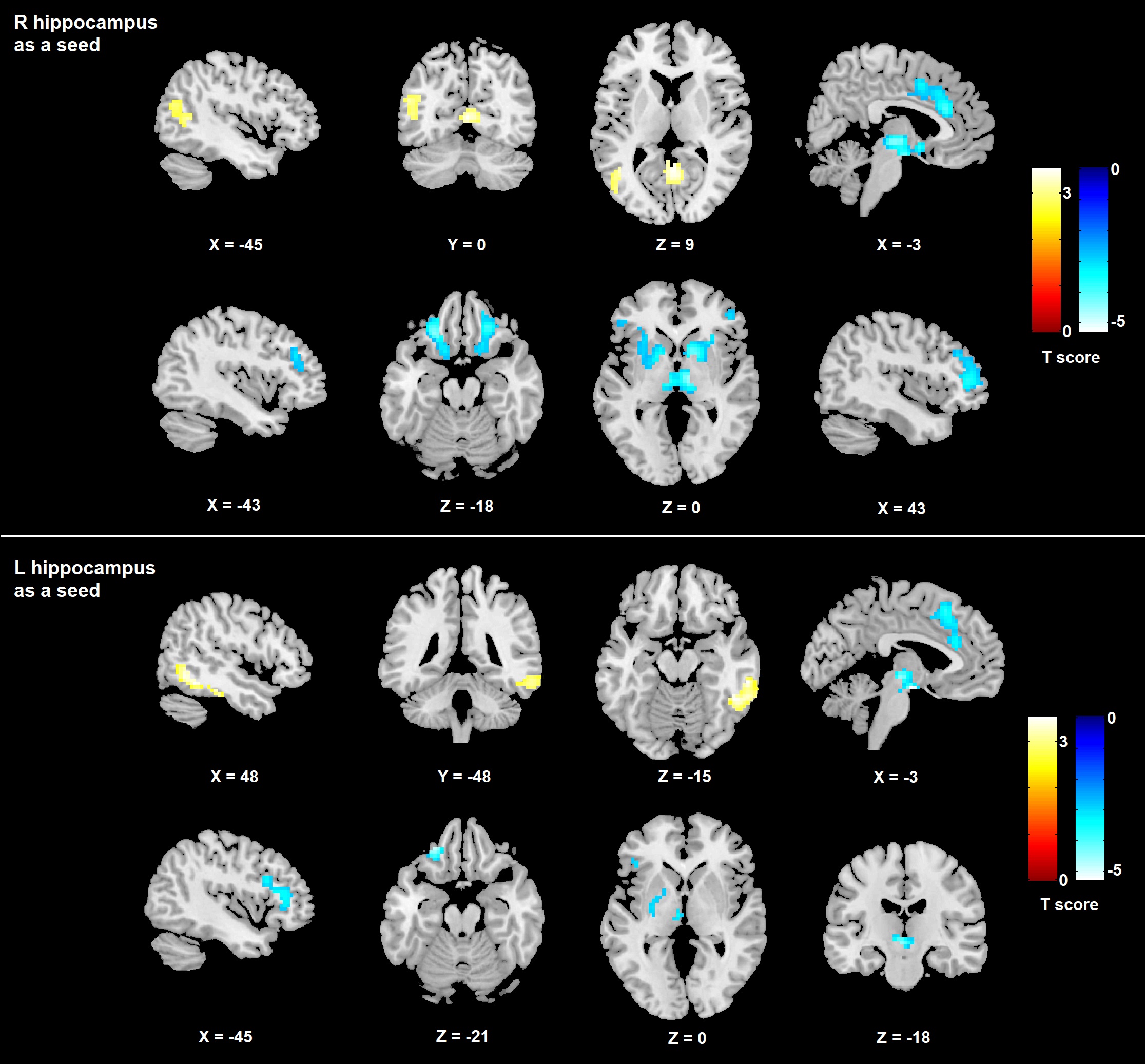
Figure 2. Significant positive (warm colors) and negative (cool colors) functional connectivity with the right and left hippocampus in first-episode drug-naïve MDD patients and HCS. Clusters are shown after controlling for multiple comparisons with AlphaSim correction (p<0.05). The color bar represents the range of T values.
Abbreviations: MDD, major depressive disorder; HCS, healthy control subjects; R, right; L, left.

Table 1. Demographic, clinical characteristics of all participants.
Abbreviations: MDD, major depressive disorder; HCS, healthy control subjects; HAMD, Hamilton Rating Scale for Depression; SD, standard deviation.

Table 2. Region-specific differences in ALFF between patients with first-episode drug-naïve major depressive disorder and healthy control subjects.
Data corrected for multiple comparisons with AlphaSim correction (p<0.05).
Abbreviations: ALFF, amplitude of low-frequency fluctuation; MDD, major depressive disorder; HCS, healthy control subjects; dACC, dorsal anterior cingulate cortex; OFC, orbitofrontal cortex; MTG, middle temporal gyrus; R, right; L, left.

Table 3. Significant differences of FC between patients with first-episode drug-naïve major depressive disorder and healthy control subjects.
Data corrected for multiple comparisons with AlphaSim correction (p<0.05).
Abbreviations: MDD, major depressive disorder; HCS, healthy control subjects; MTG, middle temporal gyrus; OFC, orbitofrontal cortex; dACC, dorsal anterior cingulate cortex; DLPFC, dorsal lateral prefrontal cortex; ITG, inferior temporal cortex; DMPFC, dorsal medial prefrontal cortex; VLPFC, ventral lateral prefrontal cortex; R, right; L, left.