2685
Structural brain abnormalities in MDD patients with suicide: A DARTEL-enhanced voxel-based morphometry study1Huaxi MR Research Center (HMRRC), Department of Radiology, West China Hospital of Sichuan University, Chengdu, China, 2Department of Psychiatry, West China Hospital of Sichuan University, Chengdu, China
Synopsis
We performed a VBM analysis with DARTEL to
analysis the different structure in healthy controls,
MDD patients with or without suicidal actors. The result shows suicidal patients had
reduced GMV than patient controls in precuneus/cuneus, anterior cingulate
cortex and orbital frontal gyrus. Particularly,
we found suicidal ideators have reduced GMV in middle frontal gyrus compared to
suicidal attempters. Negative correlation was found between clinical characters
and volume of some regions. The dysfunction of self-awareness, emotional
processing and impulsivity control function caused by the abnormalities of
these brain regions may be associated suicidal behavior.
Background
The aetiology of suicidal behavior is complex, and the depression is an
important predisposing factor to suicidal behavior. But knowledge about its
neurobiological mechanisms is limited and there is no consensus opinion on structure
MRI imaging findings of the suicidal brain of depression. This study aims to
find more detailed insight into grey matter structure and to obtain evidence
for neuroanatomical difference in suicidal brain.
Method
This study was approved by the local ethics committee, and all subjects provided written informed consent. Participant was included 47 healthy controls, 30 depressions without suicide (patient controls) and 40 depressions with suicidal behaviors (20 suicidal ideators and 20 suicidal attempters). Age and sex were matched among the three groups. Clinical symptoms were assessed with the Hamilton Depression Rating Scale (HAMD), Hamilton Anxiety Rating Scale (HAMA) and Barratt Impulsiveness Scale (BIS) among patient controls and suicidal behaviors.
We performed a voxel-based morphometry (VBM) with Diffeomorphic Anatomical Registration Through Exponentiated Lie Algebra (DARTEL) to analysis the different structure. The analysis was performed using an automated Computational Anatomy Toolbox (CAT12) within Statistical Parametric Mapping (SPM12) while running MATLAB (R2013b). T1 images were normalized to a template space and segmented into gray matter (GM), white matter (WM) and cerebrospinal fluid (CSF). And then smoothed using a 6-mm full-width-at-half-maximum (FWHM) Gaussian kernel. We using an analysis of covariance (ANCOVA) with whole-brain volume, age, and gender as covariates to compare group differences of gray matter volume (GMV) and then a subsequent post-hoc analysis used a two-sample t-test. Statistical inferences were performed with a threshold of p<0.05 with family wise error correction (FWE). Then we particularly performed a two-sample t-test between SAs and SIs.
Result
GMV across all three groups differed in bilateral precuneus, right cuneus, bilateral anterior cingulate cortex, and left superior orbital frontal gyrus. And the post-hoc t-tests showed that suicidal patients had reduced GMV than patient controls in bilateral precuneus, right cuneus, bilateral anterior cingulate cortex, and left superior orbital frontal gyrus. Moreover, we also found the patients controls showed increased GMV in bilateral precuneus and right cuneus than healthy controls. The t test analysis between SAs and SIs shows increased GMV in SAs than SIs in left middle frontal gyrus. Significant negative correlations with HAMD and HAMA scores were observed in left precuneus, and BIS scores was negatively correlated in right cuneus for GMV in suicidal groups.Discussion
The precuneus plays an important role in processing of self-relevant information, especially self-awareness1,2. High self-awareness may increase motivation to suicide3. Previous MRI studies found abnormal structure and decreased activation in precuneus/cuneus in SAs of MDD4-6. The decreased volume of precuneus and negative correlation in depression level suggest the deficits of precunes/cuneus function is associated to suicide behavior in MDD.
Both anterior cingulate cortex and orbital frontal gyrus are involved in emotional processing7, 8.Self-referential suicidal ideation such as hopelessness may be related to the alteration of ACC9. In previous studies, the alteration of structure and function of ACC were found in suicidal brain9-12. Abnormal orbitofrontal cortex makes patients more prone to making dangerous choices such as suicide13. Previous fMRI studies found in suicidal brain the OFC has reduced volume and performed less actively during decision-making some tasks13-15. Current findings of ACC and OFC are consistent with these results.
The middle frontal gyrus includes the dorsolateral prefrontal area, which plays a critical role in the development of SAs. DLPFC is a region associated with executive functions16. A systematic review shows suicidality may be associated with executive function deficits17. Previous studies found functional abnormality of the frontal cortex in SAs is related to the degree of impulsivity18, 19. Particularly, a PET study investigated rCMRglu was different in suicidal ideators and suicidal plans20. Our result may suggest MFG contributes to impulsive traits, which is associated with a vulnerability of suicidal behavior.
Conclusion
We found the GMV changes in the precuneus/cuneus, anterior cingulate cortex, orbital frontal gyrus and middle frontal gyrus in suicidal attempters. The dysfunction of self-awareness, emotional processing and impulsivity control function caused by the abnormalities of these brain regions may be associated with the vulnerability for suicidal behavior in MDD.Acknowledgements
This study was supported by the National Natural Science Foundation (Grant Nos. 81771718, 81621003, 81571637 and 81271532). Q.G. received the support from Changjiang Scholar Professorship Award (Award No. T2014190) of China and the CMB Distinguished Professorship Award (Award No. F510000/G16916411) administered by the Institute of International Education, USA.References
1. Kircher, T.T., et al., The neural correlates of intentional and incidental self processing. Neuropsychologia, 2002. 40(6): p. 683-92.
2. Kjaer, T.W., M. Nowak, and H.C. Lou, Reflective self-awareness and conscious states: PET evidence for a common midline parietofrontal core. Neuroimage, 2002. 17(2): p. 1080-6.
3. Selimbegovic, L. and A. Chatard, The mirror effect: self-awareness alone increases suicide thought accessibility. Conscious Cogn, 2013. 22(3): p. 756-64.
4. Minzenberg, M.J., et al., Control-related frontal-striatal function is associated with past suicidal ideation and behavior in patients with recent-onset psychotic major mood disorders. J Affect Disord, 2015. 188: p. 202-9.
5. Osuch, E., et al., Functional MRI of pain application in youth who engaged in repetitive non-suicidal self-injury vs. psychiatric controls. Psychiatry Res, 2014. 223(2): p. 104-12.
6. Giakoumatos, C.I., et al., Are structural brain abnormalities associated with suicidal behavior in patients with psychotic disorders? J Psychiatr Res, 2013. 47(10): p. 1389-95.
7. Allman, J.M., et al., The anterior cingulate cortex. The evolution of an interface between emotion and cognition. Ann N Y Acad Sci, 2001. 935: p. 107-17.
8. Fettes, P., L. Schulze, and J. Downar, Cortico-Striatal-Thalamic Loop Circuits of the Orbitofrontal Cortex: Promising Therapeutic Targets in Psychiatric Illness. Front Syst Neurosci, 2017. 11: p. 25.
9. Wagner, G., et al., Structural brain alterations in patients with major depressive disorder and high risk for suicide: evidence for a distinct neurobiological entity? Neuroimage, 2011. 54(2): p. 1607-14.
10. Wagner, G., et al., Prefrontal cortical thickness in depressed patients with high-risk for suicidal behavior. J Psychiatr Res, 2012. 46(11): p. 1449-55.
11. Jia, Z., et al., Impaired frontothalamic circuitry in suicidal patients with depression revealed by diffusion tensor imaging at 3.0 T. J Psychiatry Neurosci, 2014. 39(3): p. 170-7.
12. Pan, L.A., et al., Differential patterns of activity and functional connectivity in emotion processing neural circuitry to angry and happy faces in adolescents with and without suicide attempt. Psychol Med, 2013. 43(10): p. 2129-42.
13. Jollant, F., et al., Decreased activation of lateral orbitofrontal cortex during risky choices under uncertainty is associated with disadvantageous decision-making and suicidal behavior. Neuroimage, 2010. 51(3): p. 1275-81.
14. Jollant, F., et al., Orbitofrontal cortex response to angry faces in men with histories of suicide attempts. Am J Psychiatry, 2008. 165(6): p. 740-8.
15. Monkul, E.S., et al., Fronto-limbic brain structures in suicidal and non-suicidal female patients with major depressive disorder. Mol Psychiatry, 2007. 12(4): p. 360-6.
16. Forbes, C.E., et al., The role of executive function and the dorsolateral prefrontal cortex in the expression of neuroticism and conscientiousness. Soc Neurosci, 2014. 9(2): p. 139-51.
17. Bredemeier, K. and I.W. Miller, Executive function and suicidality: A systematic qualitative review. Clin Psychol Rev, 2015. 40: p. 170-83.
18. Ryding, E., et al., Regional brain serotonin and dopamine transporter binding capacity in suicide attempters relate to impulsiveness and mental energy. Psychiatry Res, 2006. 148(2-3): p. 195-203.
19. Lindstrom, M.B., et al., Impulsivity related to brain serotonin transporter binding capacity in suicide attempters. Eur Neuropsychopharmacol, 2004. 14(4): p. 295-300.
20. van Heeringen, K., et al., Decreased resting state metabolic activity in frontopolar and parietal brain regions is associated with suicide plans in depressed individuals. J Psychiatr Res, 2017. 84: p. 243-248.
Figures

Significant GMV differences are observed among healthy controls, patients controls and suicidal actors in right precuneus/cuneus (A), left orbital frontal gyrus (B) and left anterior cingulate cortex (C) shown projected onto the gray matter template built using DARTEL (corrected for multiple comparisons using a family-wise-error rate with p < 0.05). Significant GMV decrease in suicidal ideators compared with suicidal attempters in left middle frontal gyrus (D).
Abbreviation: GMV=Gray matter volume.

Both HAMD scores and HAMA scores are negatively corelated with GMV of left precuneus (A and B). BIS scores are negatively corelated with GMV of right cuneus (C).
Abbreviation: HAMD=Hamilton Depression Rating Scale; HAMA=Hamilton Anxiety Rating Scale; BIS=Barratt Impulsiveness Scale; GMV=Gray matter volume.
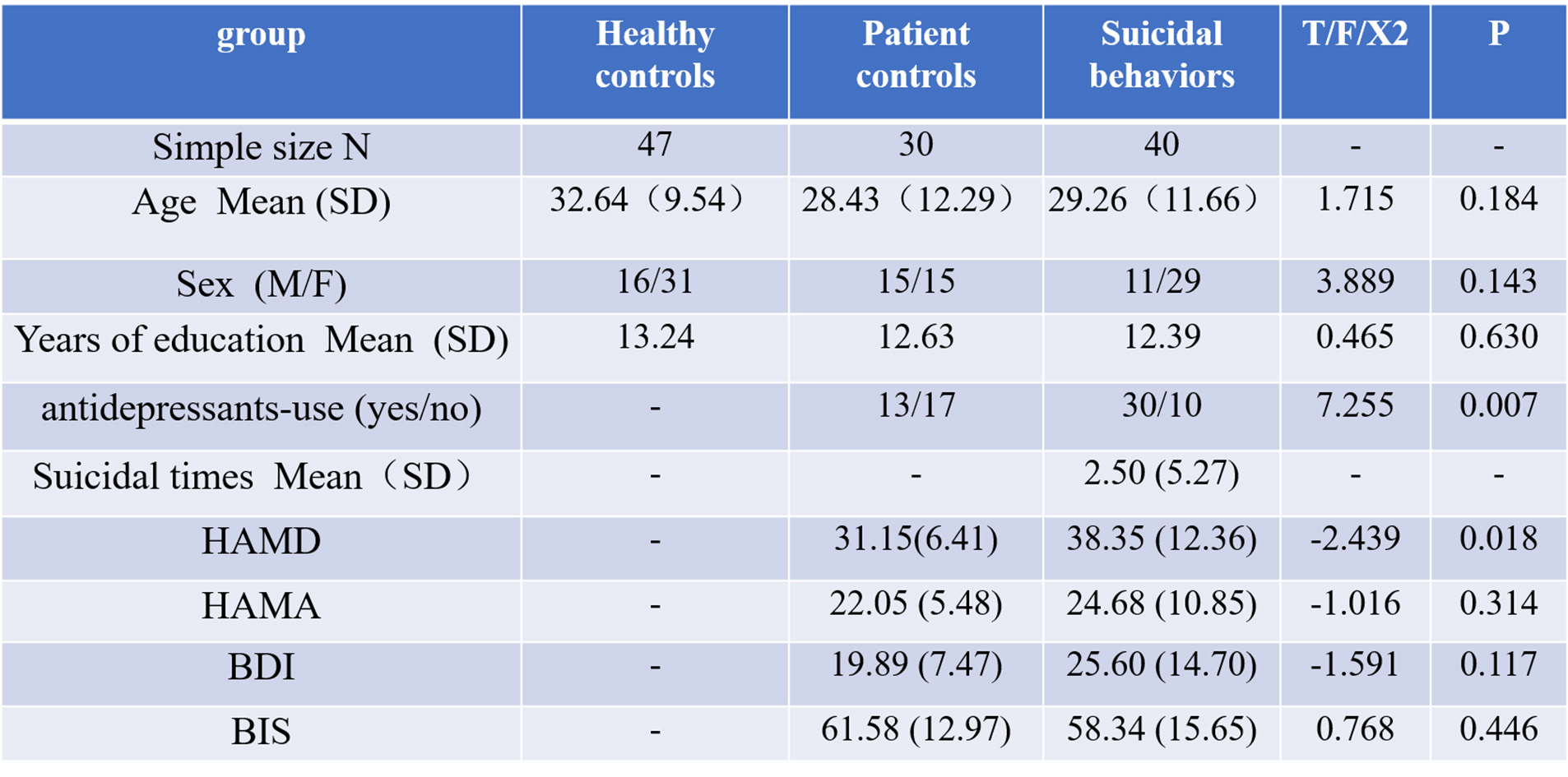
Table 1: Demographic and clinical characteristics of participants (n = 117).
Abbreviation:
HAMD=Hamilton Depression Rating Scale; HAMA=Hamilton Anxiety Rating Scale; BIS=Barratt
Impulsiveness Scale; BDI= Beck Depression Inventory.

Table 2: Demographic and clinical characteristics of suicidal actors (n = 40).
Abbreviation: HAMD=Hamilton Depression Rating Scale; HAMA=Hamilton Anxiety Rating Scale; BIS=Barratt Impulsiveness Scale; BDI= Beck Depression Inventory.

Table 3: Voxel-based analysis of gray matter volumes in MDD patients with or without suicidal history and healthy controls.
Abbreviation: SA=Suicidal attempters; SI=Suicidal ideators.