2684
Study of gender differences in major depressive disorder by using resting state brain functional magnetic resonance imaging1Radiology, West China Hospital of Sichuan University, Chengdu, China, 2The Second People’s Hospital of Yibin, Yibin, China
Synopsis
Sex differences are observed in epidemiological and clinical symptoms of major depressive disorder (MDD); yet, little is known about about the gender difference of brain function in MDD. In this work, variance analysis were used to assess the sex differences of amplitude of low frequency fluctuation (ALFF) alterations in male, female MDD patients and matched controls. We found the gender differences of ALFF in bilateral caudate nucleus and posterior cingulate gyrus. Our findings suggest that sex specific functional alterations existed in MDD, and these alterations may associated with the clinical symptoms.
Introduction
Major depressive disorder (MDD) ranks as the leading cause of years lived with disability among all diseases1. The incidence rate of men and women with MDD is about 1:2.2 The clinical manifestations of male and female patients with MDD are also different.3 However, there are few studies about the gender difference of brain function in MDD patients. Thus, the present study aimed to explore the gender differences of resting low frequency amplitude in Major depressive disorder (MDD) and the correlation between these differences and clinical manifestations.Methods
Twenty five male and 36 female unmedicated MDD patients and 25 male and 36 female age matched healthy controls were recruited in current study. The diagnosis of MDD was made using the SCID (Structured Clinical Interview for DSM Disorders) according to Diagnostic and Statistical Manual of Mental Disorders, 4th edition (DSM-IV) criteria. All subjects performed resting-state functional magnetic resonance imaging (fMRI) on 3.0T MRI (EXCITE, GE medical system, Milwaukee, WI, USA). Variance analysis was used to compare the gender differences among four groups. Correlation analysis was made between the group difference of ALFF values and the scores of Hamilton Depression Scale (HDRS) and the illness duration in MDD patients.Results
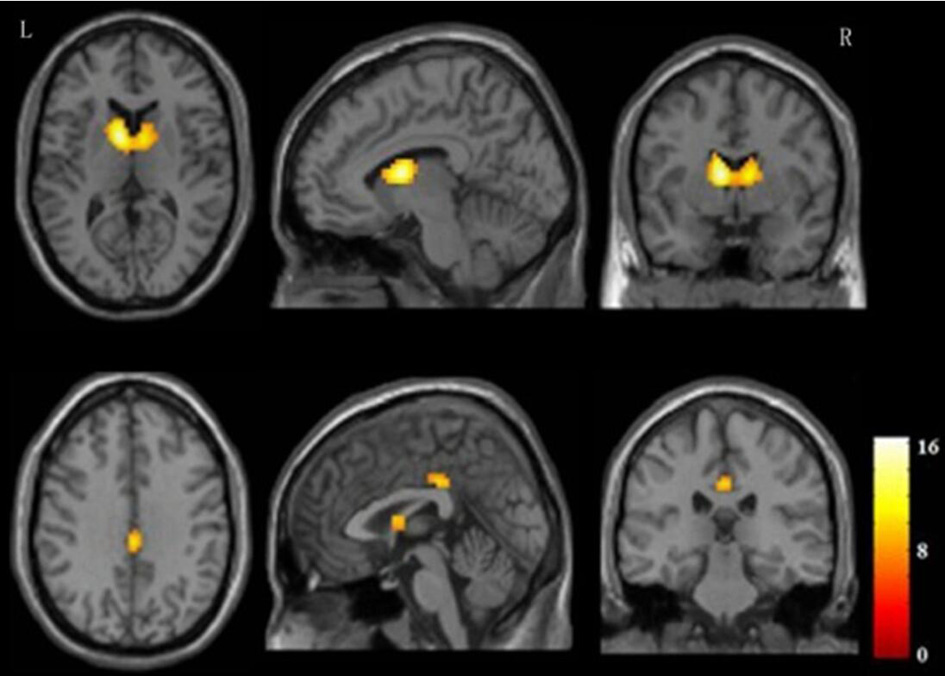
The main effect result revealed gender differences in bilateral caudate nucleus and posterior cingulate gyrus, and further post-hoc comparisons revealed the decreased ALFF of bilateral caudate nucleus and posterior cingulate gyrus mainly between male MDD patients and female MDD patients/female healthy controls (P<0.05, corrected for FWE) (Figure 1). In addition, the ALFF of bilateral caudate nucleus in female MDD patients were positively correlated with the illness duration ( r = 0.350,P=0.04 ).Discussion
Caudate nucleus, as an important part of striatum, is considered to play an important role in the occurrence and development of MDD.4 Estrogen has neuroprotective effects on the striatum, which may explain the decreased ALFF in male MDD patients in our present study.5 The positive correlation between ALFF of bilateral caudate nucleus and the illness duration in female MDD patients may be a compensatory mechanism for female patients who have the relatively short duration. Posterior cingulate gyrus, as the back node of default mode network (DMN), plays an important role in DMN. Previous study confirmed the gender differences in neurological responses to emotional stimuli in MDD patients, and male and female MDD patients have different reactive degree in the posterior cingulate cortex.6 The decreased ALFF value of the posterior cingulate gyrus suggested the nerve activity of the posterior cingulate gyrus was inhibited in male MDD which might explain why the insomnia and irritable symptoms often found in male MDD patients.Conclusion
Our findings suggest that sex specific brain functional alterations existed in MDD and the difference of ALFF in bilateral caudate nucleus and posterior cingulate gyrus between male and female MDD may be related to the different clinical manifestations in different gender of MDD.Acknowledgements
This work was supported by Sichuan Science andTechnology Program (No. 2018JY0666), 58th batches Chinese Postdoctoral ScienceFoundation (No. 2015M582554), Sichuan Provincial Health and Family Planning Commission (No. 150251) and Science and Technology Bureau of Yibin city (No.2015SF030).References
1. Moussavi S, Chatterji S, Verdes E, Tandon A, Patel V, Ustun B. Depression, chronic diseases, and decrements in health: results from the World Health Surveys. Lancet 2007; 370: 851–858.
2. Boivin JR, Piekarski DJ, Wahlberg JK, et al. Age, sex, and gonadal hormones differently influence anxiety- and depression-related behavior during puberty in mice. Psychoneuroendocrinology, 2017;85(11):78-87.
3. Khan AA, Gardner CO, Prescott CA, et al. Gender differences in the symptoms of major depression in opposite-sex dizygotic twin pairs. Am J Psychiatry, 2002,159(8):1427-1429.
4. Kim MJ, Hamilton JP, Gotlib IH. Reduced caudate gray matter volume in women with major depressive disorder. Psychiatry Res, 2008;164(2):114-122.
5. Dluzen DE. Neuroprotective effects of estrogen upon the nigrostriatal dopaminergic system. J Neurocytol, 2000;29(5-6):387-399.
6. Hofer A, Siedentopf CM, Ischebeck A, et al. Gender differences in regional cerebral activity during the perception of emotion: a functional MRI study. Neuroimage, 2006;32(2):854-862.
Figures