2683
Alterations of White Matter Tracts in Suicidal and Non-suicidal Brain with Major Depressive Disorder1Huaxi Magnetic Resonance Research Center (HMRRC), Department of Radiology, West China Hospital, Sichuan University, Chengdu, China, 2West China School of Public Health, Sichuan University, Chengdu, China
Synopsis
We investigated the white matter alterations at the individual level in MDD patients with and without suicide attempts using Automated Fiber Quantification (AFQ) approach. The three major left hemispheric white matter tracts including arcuate, CST and ATR suggested to play an important role in suicidal brain, which implies deficits of dominant hemisphere specialization with cognitive processes such as reading, writing and speaking. Our study contributes to revealing neurobiological mechanism of suicide attempts.
Background
Suicide is increasingly recognized as a concerning global social and public health problem and it is estimated that more than half of all people dying from suicide meet the criteria for major depressive disorder (MDD) [1]. However, the neurobiology of suicide in MDD patients is still largely unknown. Neuroimaging studies of the suicidal brain have characterized network abnormalities rather than deficit in a single brain region. The integrity of white matter (WM) reflects structural basis for brain functional network which can be identified by diffusion tensor imaging (DTI).
However, commonly used approaches of whole brain DTI analysis, including voxel-based analysis (VBA) and tract-based spatial statistics (TBSS), cannot accurately align fiber tracts due to variation in tract size and shape. Automated Fiber Quantification (AFQ), a new method to demonstrate the systematic variation along the trajectory of each fiber[2], provides opportunity to characterize WM microstructure alterations at the individual level. Thus, in this study we aimed to investigate the whole brain WM alterations in MDD patients with a special focus on suicide attempts using AFQ approach.
Methods
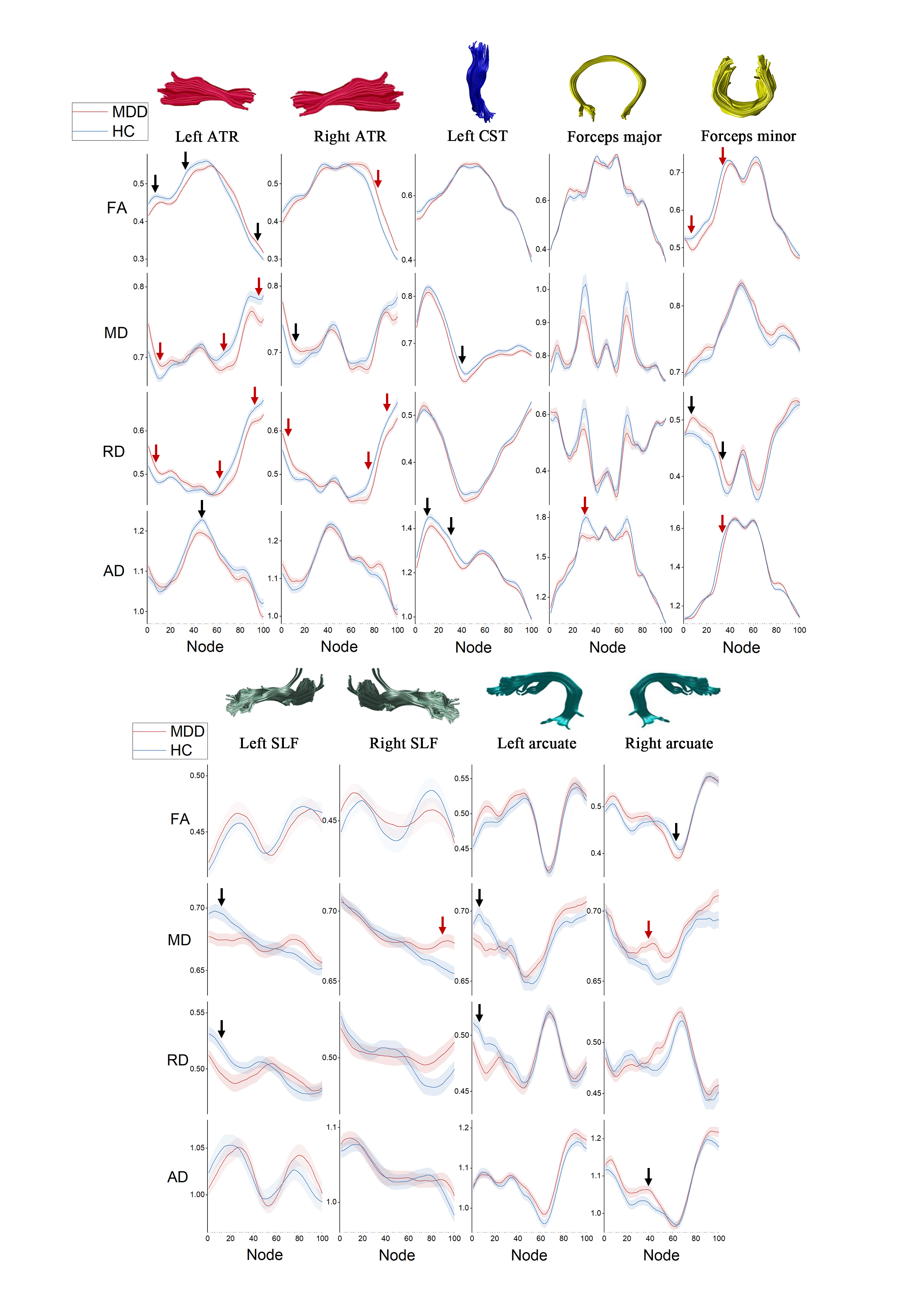
Fifty-four medication-free MDD patients with a history of suicide attempts (MDD-S) and non-suicide attempts (MDD-NS) diagnosed according DSM-IV criteria and fifty-three healthy controls (HC) were recruited. All images were acquired on a 3 T GE EXCITE MRI scanner using a single-shot echo planar imaging sequence with an 8-channel phased array head coil. The protocol included high-resolution three-dimensional T1-weighted images (TE/TR= 3.4/8.5 ms, 156 axial slices with thickness of 1 mm, axial field of view=240×240 mm, data matrix=256×256) and diffusion weighted images (TE/TR= 70.8/10,000 ms, slice thickness=3.0 mm, field of view=240×240 mm, voxel dimensions=1×1×3 mm3, scan matrix=128×128, b value=1,000 sec/mm2). Diffusion images processing was conducted using FSL (http://www.fmrib.ox.ac.uk/fsl/). Sixteen fiber tracts, including bilateral anterior thalamic radiation (ATR), bilateral corticospinal tract (CST), bilateral inferior fronto-occipital fasciculus (IFOF), bilateral inferior longitudinal fasciculus (ILF), bilateral superior longitudinal fasciculus (SLF), bilateral uncinate fasciculus (UF), bilateral arcuate and forceps major and minor, were reconstructed and quantified by the diffusion measures along the tract trajectory for each subject using the AFQ software package[2] (http://www.jasonyeatman.com/software/).
Fractional anisotropy (FA), mean diffusivity (MD), radial diffusivity (RD), axial diffusivity (AD) values of each node along the tract were compared between whole MDD patients and HC using 2-sample t-test. Furthermore, comparison of group differences between MDD-S and MDD-NS patients were performed using a general linear model (GLM) with HAMD and duration as covariates. We used false discovery rate (FDR) (p<0.05) correction for multiple comparisons. Correlations between HAMD and illness duration and diffusion parameters on significant nodes were examined using Pearson’s correlation.
Results
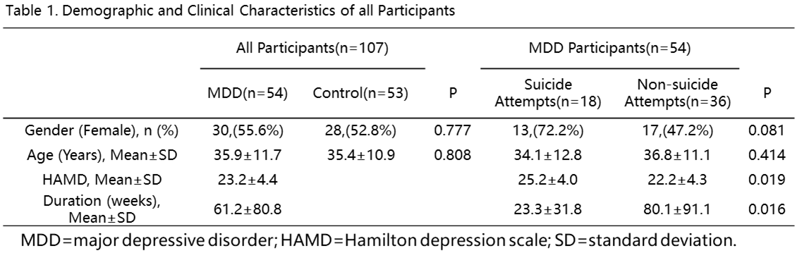
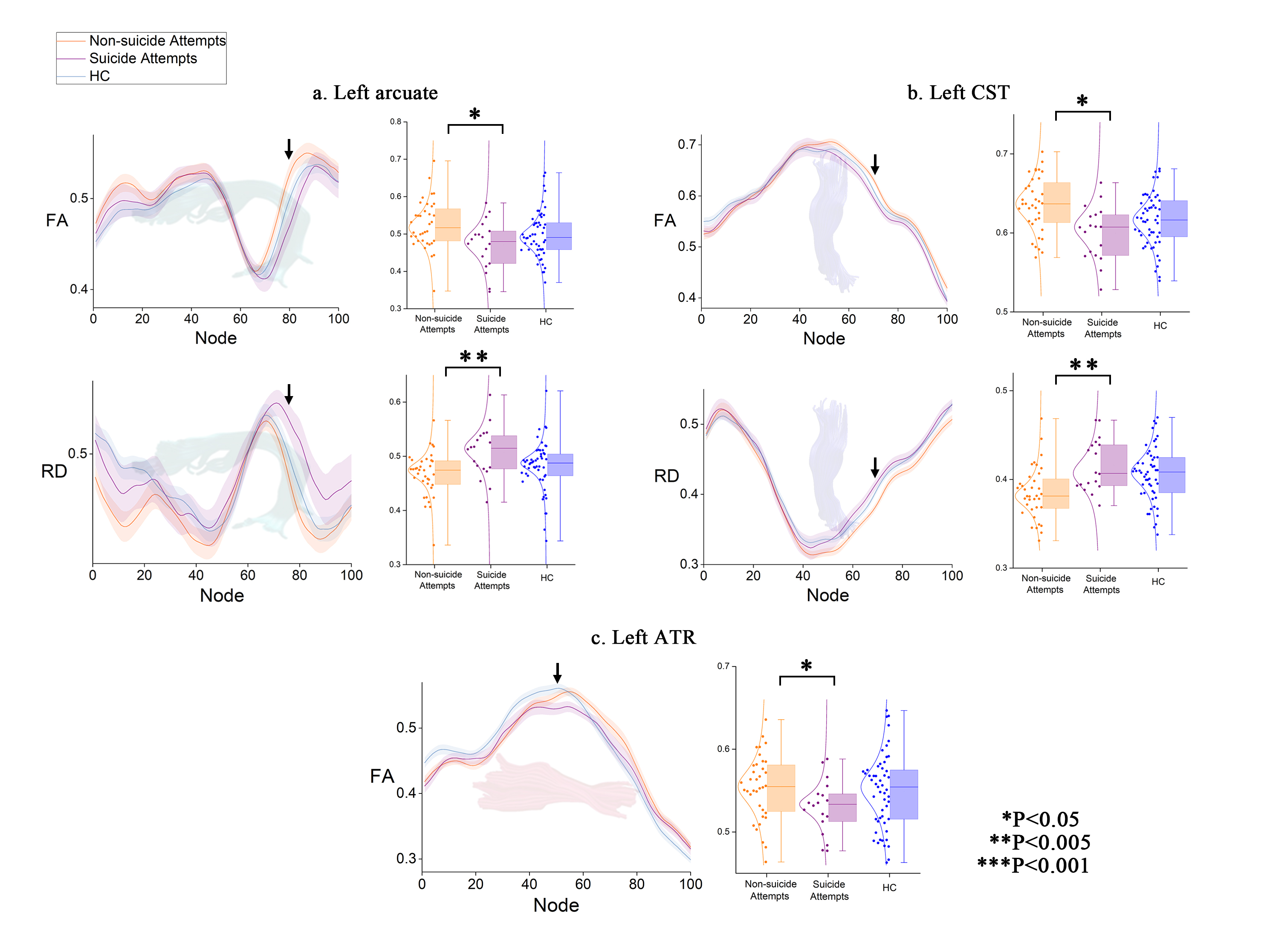
Demographic and clinical characteristics for all participants are shown in table 1. Compared with healthy controls, patients with MDD show WM alterations in bilateral ATR, left CST, forceps major and minor, bilateral SLF and bilateral arcuate (Figure 1). Furthermore, MDD-S group demonstrates significant lower FA and higher RD in the left arcuate (F=8.739, P=0.005; F=10.148, P=0.002) and left CST (F=8.008, P=0.007; F=9.573, P=0.003), lower FA in the left ATR (F=8.126, P=0.006) relative to MDD-NS group (Figure 2). No correlations between HAMD and illness duration and diffusion parameters on significant nodes were detected.Discussion and Conclusion
As far as we know, this is the first study that uses AFQ approach to demonstrate alterations of white matter tracts in suicidal brain. Three major left hemispheric white matter tracts, including arcuate, CST and ATR, are suggested to play an important role in suicidal brain. There was some evidence on hemispheric specialization with a relative left-lateralization for cognitive processes such as reading, writing and speaking, which implies deficits of cognition in suicidal brain.
We found that MDD-S present microstructural white matter alterations in the left ATR, which is consistent with our previous study[3]. However, the abnormalities of left arcuate and CST in MDD-S group are novel findings, which may attribute to the higher sensitivity of AFQ method in detecting subtle microstructural deficits in brain white matter.
Acknowledgements
This study was supported by the National Natural Science Foundation (Grant No. 81671669), Science and Technology Project of Sichuan Province (Grant No. 2017JQ0001), Undergraduate Innovation and Entrepreneurship Training Program in University of China (Grant No. C2018102362).References
1. Hawton, K. and K. van Heeringen, Suicide. Lancet, 2009. 373(9672): p. 1372-1381.
2. Yeatman, J.D., et al., Tract profiles of white matter properties: automating fiber-tract quantification. PLoS One, 2012. 7(11): p. e49790.
3. Jia, Z., et al., High-field magnetic resonance imaging of suicidality in patients with major depressive disorder. Am J Psychiatry, 2010. 167(11): p. 1381-90.
Figures