2680
Investigation of Cerebral Small Vessel Disease induced Depression using Diffusion Kurtosis Imaging - A preliminary Region-specific Study1Department of Radiology, Beijing Chaoyang Hospital (Jingxi Campus), Capital Medical University, Beijing, China, 2Department of Neurology, Beijing Chaoyang Hospital (Jingxi Campus), Capital Medical University, Beijing, China, 3Department of Radiology, Beijing Chaoyang Hospital, Capital Medical University, Beijing, China, 4MR Scientific Marketing, Siemens Healthcare, Beijing, China
Synopsis
This abstract presents a preliminary study of cerebral small vessel disease induced depression using diffusion kurtosis imaging (DKI). Different DKI-derived parameters in specific brain structures were compared between depression and non-depression groups, as well as between anxiety and non-anxiety groups. The correlation between DTI- and DKI-derived parameters and clinical scores were also investigated.
Introduction
As a group of pathological processes with various aetiologies that affect the small arteries, arterioles, venules, and capillaries of the brain, cerebral small vessel disease (SVD) is considered as one of the most common etiologies of vascular dementia and a leading cause of cognitive decline in aging brain. As a relatively homogeneous disease process, it can induce depression. Without the assumption of water molecular diffusion under a Gaussian distribution, diffusion kurtosis imaging (DKI)1,2, distinct from the conventional DTI, tries to fully utilize the MR diffusion measurements that are inherent to tissue microstructure. In this study, we investigated the correlation between DKI-derived parameters and clinical scores related to the symptom of depression.Methods
58 patients (mean age and standard deviation, 63.9±8.0 years old; age range 49-85 years old) with cerebral SVD were recruited in this study. The local ethics committee has approved this study, and written informed consent was obtained from all the patients. The severity of patient’s depression and anxiety, was evaluated for using the Hamilton Rating Scale for Depression (HRSD)3 and Hamilton Anxiety Rating Scale (HAM-A)4. T1-weighted images were acquired for anatomical reference using 3D MP-RAGE with the following parameters: TR = 2300 ms, TI = 900 ms, TE = 89 ms, FA= 8 deg, FOV =240×240 mm2, voxel size = 0.9 mm isotropic, parallel acquisition techniques (PAT) factor = 2, acquisition time = 5 min 21 sec. Diffusion-weighted images were acquired in two blocks using a spin-echo echo planar imaging (SE-EPI) pulse sequence: (i) TR = 7700 ms, TE = 89 ms, matrix = 74×74, FOV =222×222 mm2, slices = 50, slice thickness = 3 mm, no gap, b = 0, 1000, 2000 mm2/s, 1 average, 30 gradient directions, PAT factor = 2, acquisition time = 8 min 14 sec; (ii) identical as main block, expect for only b=0 mm2/s used, 9 averages, acquisition time = 1 min 34 sec. Thus, the total DWI acquisition time is 9 min 48 sec. Then the acquired DW images were provided into Diffusional Kurtosis Estimator (DKE) to reconstruct DKI maps. For the region based analysis of DKI-derived parameters, AAL atlas was applied in SPM 8 to automatically segmented brain regions, mainly in cortical sub-regions and subnuclei. The mean values of DKI-derived parameters, including kmean, kax, krad, kFA and mtk, as well as DTI-derived parameters, including dmean, dax, drad and FA, were calculated for each segmented ROIs. The nonparametric Mann-Whitney U test was used to compare mean DKI-derived parameters between depression and non-depression groups, as well as between anxiety and non-anxiety groups. Spearman correlation coefficients (r) were calculated to investigate the correlations between DKI-derived parameters and clinical scores (Hamilton depression scores and anxiety scores). P< .05 was considered statistically significant.Results
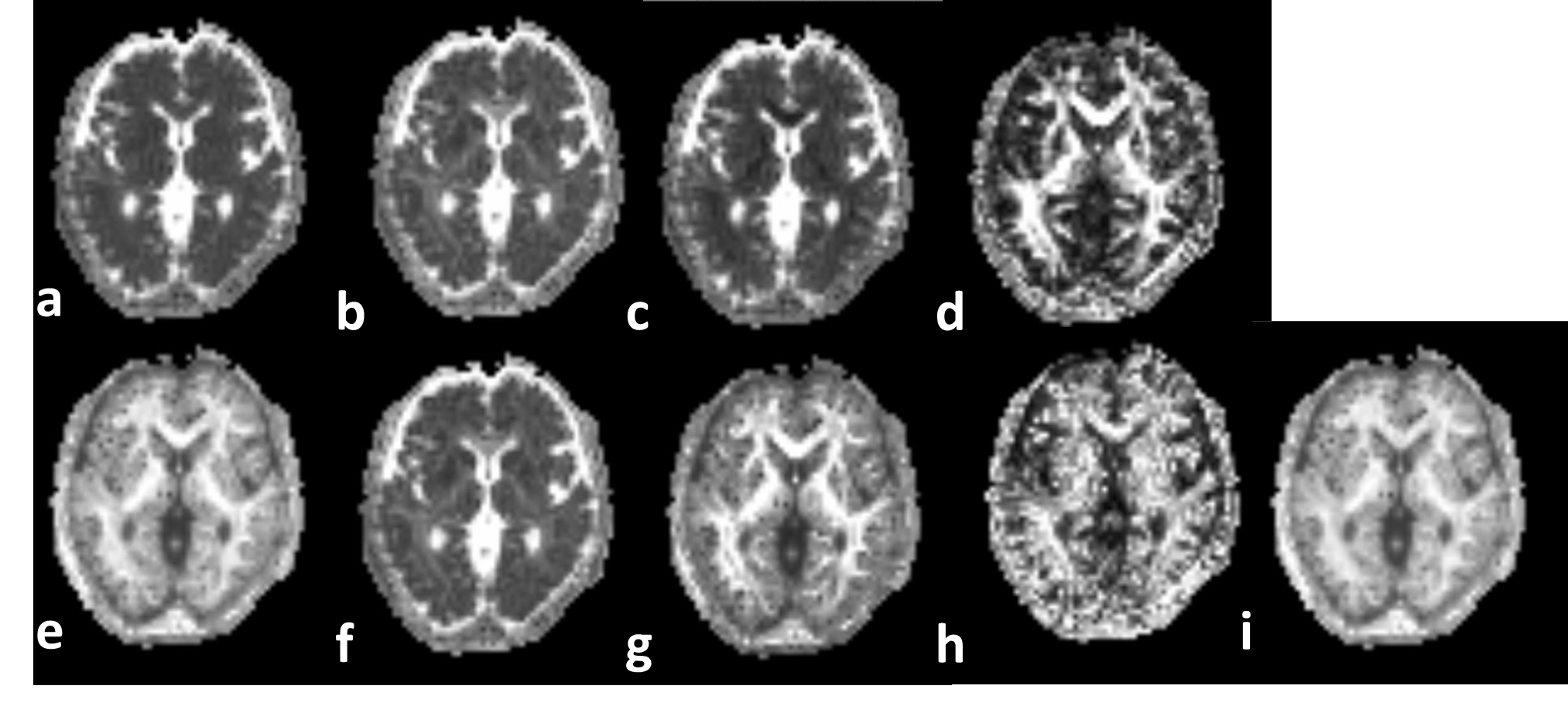
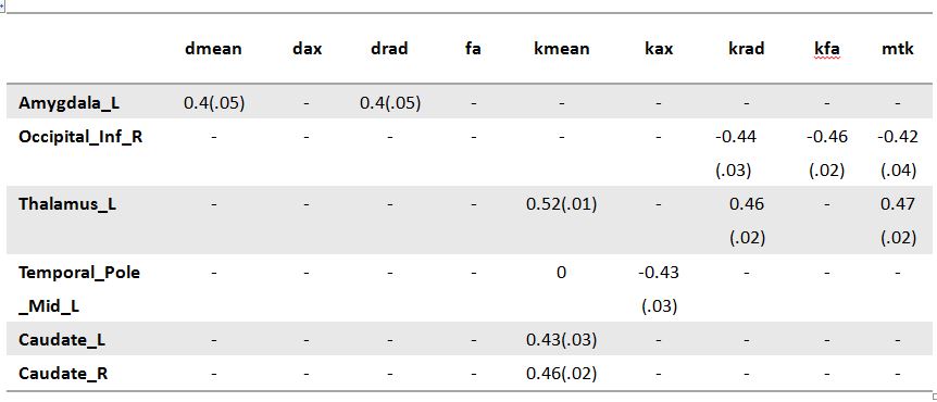
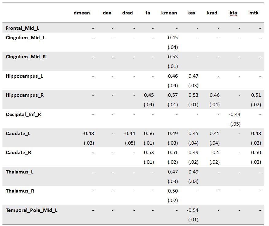
According to HRSD evaluation, 25 patients were diagnosed to be in the depression group, while the remaining 33 were put into non-depression group. According to HAM-A evaluation, 21 patients were diagnosed to have anxiety, while the remaining 37 cases had non-anxiety. Figure 1 shows an example of DTI- and DKI-derived parameters in a depressed patient. Compared to non-depression, the parameters in depression group were significantly different, including dmeam (P=.04), dax (P=.04) on left Anterior Cingulum, kmean (P=.02), mkt (P=.02) on right Anterior Cingulum, dmeam (P=.02), dax (P=.04), drad (P=.04) and kfa (P=.04) on right Amygdala, fa (P=.03), kfa (P=.02) on right middle Temporal Pole. When comparing anxiety with non-anxiety group, it turns out that fa (P=.04) on right medial orbitofrontal Cortex, dmeam (P=.03), dax (P=.04), drad (P=.04) on right anterior Cingulum, kfa (P=.04) on right thalamus were significantly different. Regarding to the correlation between DTI- and DKI-derived parameters with HRSD scores, it turns out there was significant correlation mainly on amygdala, thalamus and caudate (summarized in Table 1). While correlating to HAM-A scores, these diffusion parameters had significant correlation mainly on hippocampus, caudate, thalamus and other sub-cortex structures (summarized in Table 2).Discussion and Conclusion
In this study, it was found that DTI-derived parameters and DKI-derived parameters show substantially different characteristics on different cortical sub-regions and subnuclei when comparing depression with non-depression, anxiety and non-anxiety, as well as correlated with clinical scores. The lateral sides have also played an important role. Generally, the simple DTI model prevents it from being truly effective in characterizing relatively isotropic tissue, e.g, cortical gray matter. Instead, without assuming Gaussian distribution of water molecular diffusion, DKI might stand out when investigating the subtle changes in gray matter. The findings in this study might provide insights of the etiology, neuropathology, and pathogenesis-related microstructural and biophysical changes of depressive disorder.Acknowledgements
No acknowledgement found.References
1. Jensen JH, Helpern JA, Ramani A, et al. Diffusional kurtosis imaging: the quantification of non-gaussian water diffusion by means of magnetic resonance imaging. Magn Reson Med. 2005;53(6):1432–1440.
2. Wu EX, Cheung MM. MR diffusion kurtosis imaging for neural tissue characterization. NMR Biomed. 2010;23(7):836–48.
3. Hamilton, M. A rating scale for depression. Journal of Neurology, Neurosurgery, and Psychiatry. 1960, 23:56–62.
4. Maier W, Buller R, Philipp M, et al. The Hamilton Anxiety Scale: reliability, validity and sensitivity to change in anxiety and depressive disorders. J Affect Disord 1988;14(1):61–68.
Figures