2676
Abnormal functional connectivity of ACC sub-regions in patients with major depressive disordersXiaolong Peng1, Xiaoping Wu2, Pan Lin3, Ruxue Gong4, Rui Yang5, and Wenzhen Zhu1
1Department of Radiology, Tongji Hospital, Tongji Medical College, Huazhong University of Science and Technology, Wuhan, China, 2Department of Radiology, the Affiliated Xi'an Central Hospital of Xi'an Jiaotong University, Xi'an, China, 3Key Laboratory of Cognitive Science, College of Biomedical Engineering, South-Central University for Nationalities, Wuhan, China, 4Max Planck Institute for Human Cognitive and Brain Sciences, Leipzig, Germany, 5Department of Psychiatry, the Affiliated Xi'an Central Hospital of Xi'an Jiaotong University, Xi'an, China
Synopsis
Major depressive disorder (MDD) is a common mental disorder characterized by cognitive and affective deficits. Prior works indicated that anterior cingulate cortex (ACC) is related to high-level cognitive and emotion process, which is also thought to be pivotal to depression. Here, we examined the resting FC of ACC sub-regions in fist-episode MDD patients. The current results revealed reduced ACC sub-regional FC with IPL and SPL while increased FC was found in dmPFC. Additionally, FC with IPL also negatively correlated with symptom severity (HDRS), indicating that depression may disrupt the normal interactions within the DMN. These findings on alteration of ACC sub-regional FC may contribute to the comprehension in pathophysiology of MDD.
INTRODUCTION
Major depressive disorder (MDD) is a psychiatric disorder characterized by cognitive and affective deficits1. Anterior cingulate cortex (ACC) is considered to be involved in high-level cognitive process and emotion regulation, which is also thought to be pivotal to depression2,3. Furthermore, recent studies demonstrated that ACC could be sub-divided into different functional districts which include sensorimotor control area, cognitive process area and emotion regulation area4,5. Here, we examined the resting FC of ACC sub-regions and its relationship to symptom severity in fist-episode MDD patients.METHODS
Nineteen patients with first-episode major depressive disorders (MDD; age = 33.58 ± 9.11; 9 males) and nineteen demographic matched healthy controls (HC; age = 33.89 ± 8.88; 9 males) participated in this study. Hamilton Depression Rating Scale (HDRS) and Hamilton Anxiety Rating Scale (HARS) were applied for the behavioral assessments in MDD patients. T1-weighted images (TR = 6.9 ms; TE = 3.2 ms; flip angle = 90°; FOV = 256 mm × 256 mm; matrix = 256 ×256; slice thickness = 1.2 mm; number of slices = 128) and BOLD EPI images (TR = 2500 ms; TE = 35 ms; flip angle = 90°; FOV = 256 mm × 256 mm; matrix = 64 × 64) were collected on a 1.5-T GE Excite MRI scanner. Resting-state fMRI data were preprocessed and projected to the FreeSurfer fsaverage4 surface template using a hybrid surface- and volume-based approach. In this study, 6 seeds, named M1, C1, C2, E1, E2 and E3, were defined on each hemisphere, dividing ACC into three functional districts which include sensorimotor control area, cognitive processing area and emotional regulation area (Figure 1A). FC analysis was applied on these ACC sub-regional ROIs to all vertices on the cortical surface for each participant. Two-sample t-test (via surface-based clusterwise correction for multiple comparisons: p < 0.01, cluster size > 300 mm2) was performed to investigate the abnormal FC in MDD patients. Finally, FC of abnormal regions was compared with the symptom severity (HDRS and HDRS scores).RESULTS
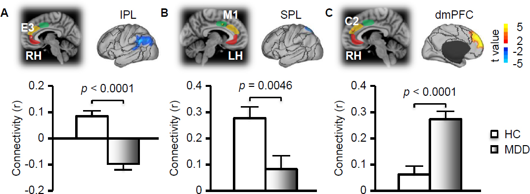
Functional connectivity of 12 ACC sub-regional seeds were computed and mean FC maps of left hemisphere seeds of MDD group were displayed in Figure 1B. Specifically, E1, E2 and E3 seeds from emotion regulation area were mainly connected with limbic regions, while C1 and C2 seeds from cognitive process area showed positive connection with medial/lateral prefrontal cortex, anterior insula and inferior parietal lobule (IPL). M1 seed mainly connected with sensorimotor cortex, superior parietal lobule (SPL) and insula. Group comparison indicated that MDD patients have reduced FC between right E3 and left IPL (p < 0.0001; Figure 2A), and between left M1 (L-M1) and left SPL (p = 0.0046; Figure 2B). Additionally, increased FC was also found in MDD patients between right C2 (R-C2) and dorsomedial prefrontal cortex (dmPFC; p < 0.0001; Figure 2C). We also found that FC between right E3 seed and left IPL negatively correlated with the improvement in HDRS scores for MDD patients (r = -0.48, p = 0.03; Figure 3).DISCUSSION
MDD patients showed reduced FC between emotion regulation area of ACC and IPL, which is involved in the perception of emotions and interpretation of sensory information. This FC also negatively correlated with HDRS scores, indicating abnormal FC alteration in IPL may be modified by depression. Besides, we found increased ACC sub-regional FC with dmPFC. Notably, ACC, IPL and dmPFC all belong to the default mode network (DMN). However, opposite connectivity changing among these regions may cause dysfunction within DMN and further disrupt the self-referential mental process, which is thought to be a potential underlying neural mechanism of depression. Additionally, as SPL is related to processing of sensory input information, reduced FC between sensorimotor areas of ACC and SPL may make patients insensitive to external stimuli and loss of interest in normal activities. These findings on disrupted FC of ACC sub-regions may contribute to understanding the pathophysiology of MDD.Acknowledgements
This work was supported by the National Natural Science Foundation of China [grant numbers 81730049; 61473221].References
1. Kessler, R. C. et al. The epidemiology of major depressive disorder: results from the National Comorbidity Survey Replication (NCS-R). Jama 289, 3095-3105 (2003). 2. Greicius, M. D. et al. Resting-state functional connectivity in major depression: abnormally increased contributions from subgenual cingulate cortex and thalamus. Biological psychiatry 62, 429-437 (2007). 3. Connolly, C. G. et al. Resting-state functional connectivity of subgenual anterior cingulate cortex in depressed adolescents. Biological psychiatry 74, 898-907 (2013). 4. Kelly, A. C. et al. Development of anterior cingulate functional connectivity from late childhood to early adulthood. Cerebral cortex 19, 640-657 (2008). 5. Margulies, D. S. et al. Mapping the functional connectivity of anterior cingulate cortex. Neuroimage 37, 579-588 (2007).Figures
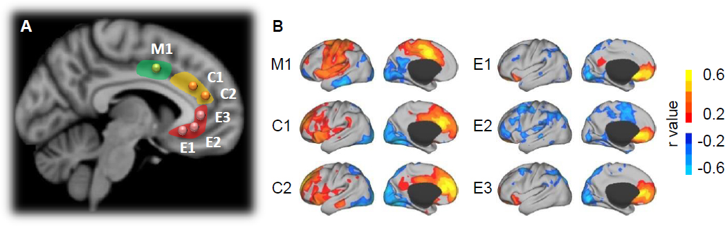
Figure 1. Functional connectivity maps of 6 left ACC sub-regions
in major depressive disorder (MDD) patients. (A) Position of ACC sub-regional
seeds on left hemisphere, include M1 from sensorimotor control area (green), C1
and C2 from cognitive process area (yellow), and E1, E2 and E3 from emotion
regulation area. (B) Mean FC maps of left ACC sub-regions in MDD patients.
Figure
2. Functional connectivity changes in patients with major depressive disorder
patients. (A) Reduced connectivity between right E3 seed and left IPL
(p<0.0001). (B) Reduced connectivity between left M1 seed and left SPL
(p=0.0046). (C) Increased connectivity between right C2 seed and left dmPFC (p<0.0001).
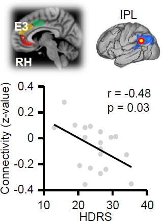
Figure
3. Hamilton depressive rate scale (HDRS) scores are significantly negative
associated with functional connectivity between right E3 seed and left IPL in
MDD patients (r=-0.48, p=0.03).