2675
Evidence for an association between low-grade peripheral inflammation and brain structural alterations in major depressionHarald Kugel1, Nils Opel2, Micah Cearns3, Scott Clark3, Catherine Toben3, Dominik Grotegerd2, Walter Heindel1, Anja Teuber4, Heike Minnerup4, Matthias Nauck5, Klaus Berger4, Udo Dannlowski2, and Bernhard T. Baune6
1Institute of Clinical Radiology, University of Münster, Münster, Germany, 2Department of Psychiatry, University of Münster, Münster, Germany, 3Discipline of Psychiatry, University of Adelaide, Adelaide, Australia, 4Institute of Epidemiology and Social Medicine, University of Münster, Münster, Germany, 5Institute of Laboratory Medicine, University of Greifswald, Greifswald, Germany, 6Department of Psychiatry, University of Melbourne, Melbourne, Australia
Synopsis
Preliminary research suggests that major depressive disorder (MDD) is associated with structural alterations of brain regions relevant for emotion regulation and associated with low-grade peripheral inflammation as indicated by high sensitive C-reactive protein (hsCRP) serum levels. This association between structural brain alterations and low-grade inflammation as potentially interrelated biological correlates of MDD was investigated. In MDD patients, but not healthy controls, prefrontal gray matter volume reductions were significantly associated with higher hsCRP levels.
Introduction
Preliminary research suggests that major depressive disorder (MDD) is associated with structural alterations of brain regions that play a role in reward processing and emotion regulation1,2 as well as with low-grade peripheral inflammation as indicated by high sensitive C-reactive protein (hsCRP) serum levels.3,4 However, even though a link between inflammatory processes and altered brain structural integrity has been purported by experimental research5,6, the biological processes underlying the associations between MDD and brain structural alterations are still poorly understood. An important question in this context is, if chronic low-grade peripheral inflammation might contribute to structural abnormalities in MDD. Well powered studies have been lacking to confirm this hypothesis in MDD patients. Therefore, we aimed to investigate the potential association between structural brain alterations and low-grade inflammation as potentially interrelated biological correlates of MDD.Methods
In this cross-sectional study 514 MDD patients and 359 healthy controls (HC) underwent structural MRI. Voxel-based-morphometry (VBM) was used to study local differences in gray matter volume. Serum levels of high sensitive C-reactive protein (hsCRP) were assessed in each participant. Due to the not normal distribution of hsCRP, logarithmic transformed hsCRP values were used for analyses. MRI images were obtained at 3 T (Gyroscan Intera with Achieva upgrade) using a 3D T1w TFE-sequence (TR/TE/FA = 7.4 ms/3.4 ms/9°, inversion prepulse every 814.5 ms, reconstructed to voxels of .5 mm edge length). Images were preprocessed using the pipeline of the CAT12-toolbox.7 Imaging analyses were done in SPM12.8 Further statistical analyses were performed with SPSS version 25. (1) Group differences in hsCRP and log hsCRP levels between MDD and HC subjects were investigated. (2) Whole brain VBM analysis was done to investigate group differences in gray matter volume between MDD and HC subjects. (3) A multiple regression analysis of log hsCRP levels on gray matter volume was performed in the MDD group, on ROIs including the significant clusters determined in the preceding (step (2)) whole brain VBM analysis. (4) A regression analysis analog to (3) was performed in the HC group. Contributions of further potentially confounding variables were checked.Results
(1) Compared to healthy controls, MDD patients exhibited significantly increased hsCRP and log hsCRP levels (p < .001). (2) MDD patients showed significantly decreased gray matter volume in the prefrontal cortex, temporal cortex, and the insula. (3) In MDD patients, prefrontal gray matter volume reductions were significantly associated with higher hsCRP levels (Fig. 1). The significant negative association between hsCRP and gray matter appeared independent of age, sex, body-mass-index, antidepressant load, hospitalization and medical comorbidities in the MDD sample. (4) In the HC group, no significant association between hsCRP levels and gray matter could be detected.Discussion
The results give evidence for a relationship between peripheral low-grade inflammation and gray matter reductions in the prefrontal cortex in MDD. In line with our hypothesis and previous reports, MDD patients showed significantly altered brain structural integrity compared to HC.2,9 Most importantly, gray matter decrease in MDD is associated with higher level of the proinflammatory marker hsCRP, while no such association is present in the HC group. The inverse association between hsCRP and gray matter was regionally specific for the prefrontal cortex, a brain region associated with MDD.1,9 Recent evidence hints to a bidirectional communication between CNS and immune system - a pro-inflammatory environment may impair processes to maintain the integrity of gray matter in the prefrontal cortex, a brain region with pronounced structural plasticity and vulnerability to environmental influences.10 In addition, the CNS is thought to exert an influence on peripheral inflammatory processes, e.g. via activation of stress signaling pathways in response to external stress.11 Considering this bidirectional communication, the MDD-specific structural alterations might in turn contribute to the maintenance of low-grade inflammation in MDD.Conclusions
The present study highlights the role of reduced gray matter volume and low-grade peripheral inflammation as interrelated biological correlates of MDD. The reported inverse association between peripheral low-grade inflammation and brain structural integrity in depressive patients translates current knowledge from experimental studies to the bedside in MDD.Acknowledgements
Funded by the German Federal Ministry of Education and Research (BiDirect study, grants 01ER0816, 01ER1506 and 01ER1205). Additional funding: German Research Foundation (DFG, grant FOR2107 DA1151/5-1 and DA1151/5-2 ; SFB-TRR58, Projects C09 and Z02 ) and Interdisciplinary Center for Clinical Research (IZKF) of the medical faculty of Münster (grant Dan3/012/17) and Deanery of the Medical Faculty of the University of Münster.References
1. Schmaal L, Hibar DP, Sämann PG, et al. Cortical abnormalities in adults and adolescents with major depression based on brain scans from 20 cohorts worldwide in the ENIGMA Major Depressive Disorder Working Group. Mol Psychiatry. 2017;22(6):900-909. 2. Schmaal L, Veltman DJ, van Erp TGM, et al. Subcortical brain alterations in major depressive disorder: findings from the ENIGMA Major Depressive Disorder working group. Mol Psychiatry. 2015;21(6):806-812. 3. Haapakoski R, Mathieu J, Ebmeier KP, et al. Cumulative meta-analysis of interleukins 6 and 1β, tumour necrosis factor α and C-reactive protein in patients with major depressive disorder. Brain Behav Immun. 2015;49:206-215. 4. Horn SR, Long MM, Nelson BW, et al. Replication and reproducibility issues in the relationship between C-reactive protein and depression: A systematic review and focused meta-analysis. Brain Behav Immun. 2018; 73:85-114. 5. Chesnokova V, Pechnick RN, Wawrowsky K. Chronic peripheral inflammation, hippocampal neurogenesis, and behavior. Brain Behav Immun 2016;58:1-8. 6. Harrison NA. Brain Structures Implicated in Inflammation-Associated Depression. Curr Top Behav Neurosci. 2017;31:221-248. 7. www.neuro.uni-jena.de/cat, Version 933. 8. www.fil.ion.ucl.ac.uk/spm, Version 6685. 9. Price JL, Drevets WC. Neural circuits underlying the pathophysiology of mood disorders. Trends Cogn Sci. 2012;16(1):61-71. 10. McEwen BS. Protection and damage from acute and chronic stress: allostasis and allostatic overload and relevance to the pathophysiology of psychiatric disorders. Ann N Y Acad Sci. 2004;1032:1-7. 11. Irwin MR, Cole SW. Reciprocal regulation of the neural and innate immune systems. Nat Rev Immunol 2011;11(9):625-632.Figures
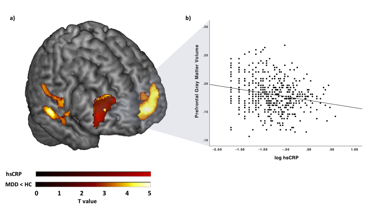
Figure 1. a) Association between log hsCRP values and MDD related gray matter: Results for the log hsCRP analysis in red color, the employed mask comprising significant clusters of the preceding whole brain analyses of group differences between HC and MDD subjects in yellow color. For display reasons uncorrected results at voxel-threshold p < 0.01, minimum cluster volume threshold k≥ 400 are presented.b) Plot depicting the negative association between log hsCRP and gray matter volume x= 50, y= 50, z= 8. Abbreviations: HC, Healthy controls; MDD, MDD patients; log hsCRP, logarithmic transformed high-sensitive C-reactive protein