2670
Changes to Blood-Brain Barrier Water Permeability After CPAP Treatment in Patients with Obstructive Sleep Apnea1Radiology, Stony Brook University, Stony Brook, NY, United States, 2Biomedical Engineering, Stony Brook University, Stony Brook, NY, United States, 3Medicine, Stony Brook University, Stony Brook, NY, United States
Synopsis
In patients with obstructive sleep apnea (OSA), intermittent ischemia and re-oxygenation leads to disruption of blood-brain barrier (BBB) integrity. In this study changes in BBB water permeability parameters, water extraction fraction (Ew) and water permeability surface area product (PSw), in patients with OSA before and after 8-weeks of continuous positive airway pressure (CPAP) treatment were investigated using the recently developed Intrinsic Diffusivity Encoding of Arterial Labeled Spins (IDEALS) technique. Compared to healthy controls, OSA patients exhibited lower CBF, PSw and Ew before CPAP. After 8-weeks of CPAP, patients showed increased CBF, PSw and Ew demonstrating the improvement of BBB integrity.
Introduction
Obstructive sleep apnea (OSA) is characterized by transient hypoxia with reduced cerebral blood flow (CBF)1 followed by re-oxygenation and elevated CBF at the event’s termination, that occur during sleep. Continuous positive airway pressure (CPAP) treatment has demonstrated beneficial effects in most studies. In OSA, intermittent ischemia and re-oxygenation leads to disruption of blood-brain barrier (BBB) integrity3-6. Because trans-capillary water exchange is mainly facilitated by active transport mechanisms7-9, assessing BBB water permeability, i.e. water extraction fraction (Ew) and water permeability surface area product (PSw), could provide a direct and sensitive assessment of subtle BBB disruption. In this study we investigate changes in BBB water permeability in patients with OSA before and after CPAP using the recently developed Intrinsic Diffusivity Encoding of Arterial Labeled Spins (IDEALS) technique10,11.Methods
Patients with moderate to severe OSA were recruited with IRB approval and informed written consent. Currently, two OSA patients (2 males; 45 ± 14 years old) completed the 8-week CPAP treatment with more patients expected to complete soon. Results from 4 age and gender matched healthy controls (4 males; 41 ± 8 years old) were included from a previous study10. All studies were performed on a Siemens 3T Biograph mMR with 12-channel head/neck coil. In the IDEALS paradigm, intravascular and extravascular water in an arterial spin labeling (ASL) experiment are separated by their different diffusion sensitivities at two segmentation factors in 3D-GRASE acquisition12. The MRI parameters were: TR/TE/Label Time 4500/16.12/1600 ms, FA=120°, matrix of 64×64×32, FOV of 256×256×128mm3, iPAT2. Two segmentation schemes (4PAR×2PE and 1PAR×2PE) and two post labeling delays (PLDs) (1000 and 2000 ms) were used for a total of 4 sets of ASL images with total acquisition time ~15 min. The arterial transit time was estimated from the 1000 ms PLD data. CBF, Ew, and PSw were subsequently estimated from the 2000 ms PLD data. High resolution MPRAGE images were acquired for segmentation and spatial normalization. Parameter values in the following regions were evaluated: global gray matter (GM), posterior cingulate cortex (PCC), hippocampus, inferior parietal cortex (IPC), medial prefrontal cortex (MPFC), and mediotemporal lobe (MTL)13.Results
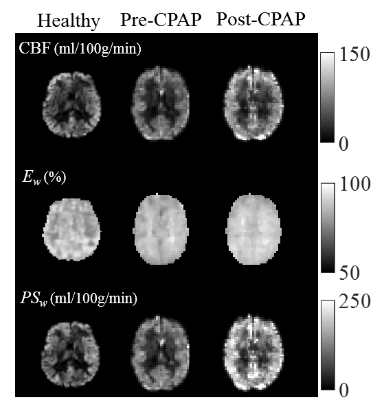
Figure 1 displays the pre- and post-treatment IDEALS parameter maps for an OSA patient (male; 35 years old) and baseline measurement from a healthy control (male; 34 years old). Compared to healthy controls, OSA patients exhibited lower baseline global GM CBF (45.3 ± 3.0 vs. 57.2 ± 8.5 mL/100g/min), Ew (83.4% ± 1.5% vs. 84.0% ± 1.8%) and PSw (86.0 ± 6.9 mL/100g/min vs. 102.3 ± 16.4 mL/100g/min). OSA patients demonstrated marked increases of CBF, Ew, and PSw after 8-weeks of compliant CPAP treatment. Increases to global GM CBF (58.1 ± 5.0 vs. 45.3 ± 3.0 mL/100g/min), Ew (85.2% ± 0.9% vs. 83.4% ± 1.5%) and PSw (115.7 ± 11.1 vs. 86.0 ± 6.9 mL/100g/min) were also observed. In addition to global changes, regional after/before changes were similar: increased CBF in the PCC (56.0 ± 10.3 vs. 55.2 ± 12.0 mL/100g/min), hippocampus (40.7 ± 8.0 vs. 38.5 ± 6.2 mL/100g/min), IPC (60.7 ± 11.9 vs. 56.2 ± 5.4 mL/100g/min), MPFC (69.8 ± 18.5 vs. 51.5 ± 15.7 mL/100g/min), and MTL (72.1 ± 18.9 vs. 55.0 ± 10.5 mL/100g/min); increased Ew in the PCC (89.4% ± 2.1% vs. 87.3% ± 3.6%), hippocampus (86.3% ± 2.4% vs. 84.8% ± 3.4%), IPC (87.2% ± 1.8% vs. 85.6% ± 0.9%), MPFC (89.3% ± 1.6% vs. 86.8% ± 4.0%), and MTL (88.0% ± 3.3% vs. 86.4% ± 3.3%); increased PSw in the PCC (147.4 ± 36.0 vs. 113.2 ± 8.3 mL/100g/min), hippocampus (81.5 ± 18.0 vs. 74.0 ± 10.1 mL/100g/min), IPC (124.6 ± 24.7 vs. 110.5 ± 11.9 mL/100g/min), MPFC (156.6 ± 30.2 vs. 105.6 ± 18.0 mL/100g/min), and MTL (155.5 ± 41.6 vs. 111.7 ± 12.9 mL/100g/min).Discussion
This study observed lower baseline BBB water permeability in OSA patients compared to healthy controls and an increase in BBB water permeability in OSA patients after 8-weeks of CPAP treatment. The lower BBB water permeability at baseline demonstrates disruption of BBB integrity due to OSA3-6. With active transmembrane water cycling pathways, such as NaK-ATPase, accounting for a large fraction of water exchange14,15, the increase of BBB water permeability observed after treatment in OSA patients suggests normalization of BBB integrity and cerebral metabolism16. These improvements were observed in several GM regions implicated in OSA13.Conclusion
Changes to BBB water permeability in patients with OSA in response to 8-weeks of compliant CPAP treatment were measured using the IDEALS approach. Increases to CBF, Ew, and PSw were observed after treatment, suggesting normalization of these parameters in response to treatment.Acknowledgements
No acknowledgement found.References
1. Jennum P, Borgesen SE. Intracranial pressure and obstructive sleep apnea. Chest. 1989;95(2):279-283.
2. Ferini-Strambi L, Baietto C, Di Gioia MR, et al. Cognitive dysfunction in patients with obstructive sleep apnea (OSA): partial reversibility after continuous positive airway pressure (CPAP). Brain research bulletin. 2003;61(1):87-92.
3. He J, Hsuchou H, He Y, Kastin AJ, Wang Y, Pan W. Sleep restriction impairs blood-brain barrier function. J Neurosci. 2014;34(44):14697-14706.
4. Busch DR, Lynch JM, Winters ME, et al. Cerebral Blood Flow Response to Hypercapnia in Children with Obstructive Sleep Apnea Syndrome. Sleep. 2016;39(1):209-216.
5. Urbano F, Roux F, Schindler J, Mohsenin V. Impaired cerebral autoregulation in obstructive sleep apnea. J Appl Physiol (1985). 2008;105(6):1852-1857.
6. Cramer SP, Simonsen H, Frederiksen JL, Rostrup E, Larsson HB. Abnormal blood-brain barrier permeability in normal appearing white matter in multiple sclerosis investigated by MRI. NeuroImage Clinical. 2014;4:182-189.
7. Loo DD, Zeuthen T, Chandy G, Wright EM. Cotransport of water by the Na+/glucose cotransporter. Proceedings of the National Academy of Sciences. 1996;93(23):13367-13370.
8. Cong D, Zhu W, S Kuo J, Hu S, Sun D. Ion transporters in brain tumors. Current medicinal chemistry. 2015;22(10):1171-1181.
9. Zhang Y, Poirier-Quinot M, Springer CS, Balschi JA. Active trans-plasma membrane water cycling in yeast is revealed by NMR. Biophysical journal. 2011;101(11):2833-2842.
10. He X, Wengler K, Duong T, Schweitzer M. 3D MRI Mapping of Whole-Brain Water Permeability with Intrinsic Diffusivity Encoding of Arterial Labeled Spins (IDEALS). In Proceedings of the 27th Annual Meeting of ISMRM, Paris. 2018;0180.
11. Wengler K, Ha J, Coyle P, Schweitzer M, Duong T, He X. Blood Brain Barrier Water Permeability in Non-Enhancing Multiple Sclerosis Lesion with Intrinsic Diffusivity Encoding of Arterial Labeled Spins (IDEALS). In Proceedings of the 27th Annual Meeting of ISMRM, Paris. 2018;4921.
12. He X, Wengler K, Schweitzer ME. Diffusion sensitivity of 3D‐GRASE in arterial spin labeling perfusion. Magnetic resonance in medicine. 2018;80(2):736-747.
13. Li H-J, Nie X, Gong H-H, Zhang W, Nie S, Peng D-C. Abnormal resting-state functional connectivity within the default mode network subregions in male patients with obstructive sleep apnea. Neuropsychiatric disease and treatment. 2016;12:203.
14. Bai R, Springer Jr CS, Plenz D, Basser PJ. Fast, Na+/K+ pump driven, steady‐state transcytolemmal water exchange in neuronal tissue: A study of rat brain cortical cultures. Magnetic resonance in medicine. 2018;79(6):3207-3217.
15. Bai R, Springer Jr CS, Plenz D, Basser PJ. Brain active transmembrane water cycling measured by MR is associated with neuronal activity. Magnetic resonance in medicine. 2018.
16. Rooney WD, Li X, Sammi MK, Bourdette DN, Neuwelt EA, Springer CS. Mapping human brain capillary water lifetime: high‐resolution metabolic neuroimaging. NMR in Biomedicine. 2015;28(6):607-623.
Figures