2668
Correlation of cerebrovascular reserve assessed by acetazolamide-stress SPECT with collaterals on arterial spin-labeling MRI in patients with carotid occlusive disease1Seoul Veterans Hospital, Seoul, Korea, Republic of
Synopsis
We evaluated the correlation between cerebrovascular reserve (CVR) on acetazolamide (ACZ) -stress SPECT brain scans and collaterals on ASL MRI in ICA stenosis. 86 patients with ICA stenosis (>70%) were enrolled in this study. On ASL, late-arriving flow appears as serpiginous high ASL signal within cortical vessels, which has been termed arterial transit artifact (ATA). 82/86 ICA stenosis patients underwent SPECT imagings with Tc-99m-ECD in the resting and after ACZ challenge. Significant positive relationship was observed between normal CVR group and ATA showing group in ICA stenosis patients on ASL brain perfusion (p=0.035, chi-square test).
Purpose
We evaluated the correlation between cerebrovascular reserve (CVR) on acetazolamide (ACZ) -stress single photon emission computed tomography (SPECT) brain scans and collaterals on arterial spin-labeling (ASL) magnetic resonance imaging (MRI) in internal carotid artery (ICA) stenosis.Methods
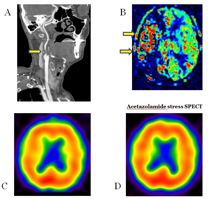
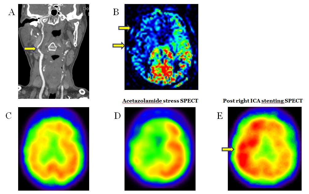
86 patients with ICA stenosis (>70%) were enrolled in this study. Including pulsed ASL, MRI was acquired on a 3 tesla system. On ASL, late-arriving flow appears as serpiginous high ASL signal within cortical vessels, which has been termed arterial transit artifact (ATA). Images were interpreted for the presence of ATA. 82/86 ICA stenosis patients underwent SPECT imagings with Tc-99m-ECD in the resting and after ACZ challenge. We observed the presence of intracranial collaterals, which are manifested by ATA, on ASL brain perfusion scan. CVR based on rest-SPECT and ACZ-stress SPECT was calculated. With ACZ-stress SPECT, the 82 patients were grouped as either showing or not showing evidence of decreased CVR. We assessed the relationship between reduced CVR and intracranial collaterals shown as ATA on ASL brain perfusion.Results
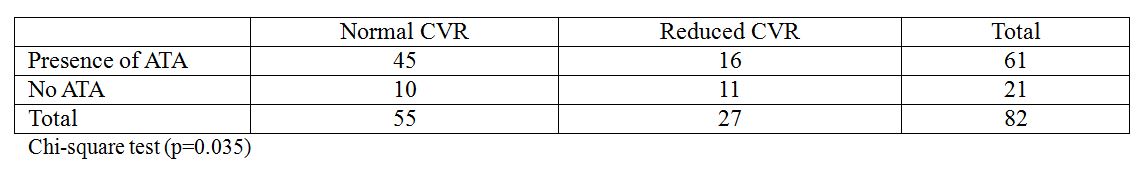
In 61/86 (70%) of the ICA stenosis patients, ASL showed ATA in ipsilateral to the stenosis. With acetazolamide stress SPECT, the 27/82 (32%) patients showed evidence of decreased CVR. In 45/55 (81%) of the normal CVR group and 16/27 (59%) of the reduced CVR from the SPECT results, pulsed ASL showed ATA in ipsilateral to the stenosis. Significant positive relationship was observed between normal CVR group and ATA showing group in ICA stenosis patients on ASL brain perfusion (p=0.035, chi-square test).Discussion
The most important finding of our study was that there is a statistically significant positive correlation is noted between normal CVR group and ICA stenosis patients with ATA. Our results are comparable to those of prior stroke study suggested that patients with ATA had improved outcomes, and ATA may represent collateral flow 1,2. And other prior study showed that the presence of ATA in patients with carotid disease was predictive of poor cerebrovascular reactivity following an ACZ challenge 3. Smith et al 4, who found an increased number of leptomeningeal collaterals in patients with a steal response to ACZ, and reported good correlation between CVR measured with ACZ-challenged xenon-enhanced CT and leptomeningeal collateralization at cerebral angiography. So, reduced CVR is significantly associated with a dependence on leptomeningeal collaterals that are presented by ATA on ASL, and implies a state of maximal hemodynamic compromise in the setting of chronic stenosis or occlusion of the ICA.Positron emission tomography (PET) and SPECT can be combined with a vasodilatory stimulus to determine the resilience of the cerebral circulation to ischemic insults, termed the CVR. Intravenous ACZ or inhaled CO2 are the most common vasodilatory stimulus, and an inability to recruit additional blood flow in response to their administration signified impaired collateral flow and diminished CVR. ASL is an MRI technique for measuring CBF at the brain tissue level. It uses radiofrequency pulses to noninvasively label water protons in blood. Previous studies have shown good correlation with gold standard CBF imaging of gray matter in healthy subjects 4, but it is likely that it underestimates CBF in regions with delayed arterial arrival times. This is because the label decays with the blood T1. However, this drawback for quantitation may be turned to advantage for visualizing collaterals. With ASL, late-arriving flow appears as serpiginous high ASL signal within cortical vessels, which has been termed ATA 5. ATA was seen frequently in a small group of acute ischemic stroke patients and was associated with tissue survival and improved clinical outcome. Also, patients with chronic hypoperfusion with ATA had good CVR in response to ACZ 6.Conclusion
The ATA with ASL imaging as a noninvasive and no contrast demanding technique, can depict slow flow in excellent collateral vessels and has clinical utility in detecting CVR in patients with ICA stenosis.Acknowledgements
No acknowledgement found.References
1. Chng SM, Petersen ET, Zimine I, Sitoh YY, Lim CC, Golay X. Territorial arterial spin labeling in the assessment of collateral circulation: comparison with digital subtraction angiography. Stroke. 2008; 39:3248–3254.
2. Chalela JA, Alsop DC, Gonzalez-Atavales JB, Maldjian JA, Kasner SE, Detre JA. Magnetic resonance perfusion imaging in acute ischemic stroke using continuous arterial spin labeling. Stroke. 2000; 31:680–687.
3. Detre JA, Samuels OB, Alsop DC, Gonzalez-At JB, Kasner SE, Raps EC. Noninvasive magnetic resonance imaging evaluation of cerebral blood flow with acetazolamide challenge in patients with cerebrovascular stenosis. J Magn Reson Imaging. 1999; 10:870–875.
4. Smith HA, Thompson-Dobkin J, Yonas H, Flint E. Correlation of xenon-enhanced computed tomography-defined cerebral blood flow reactivity and collateral flow patterns. Stroke 1994; 25:1784–1787.
5. Zaharchuk G, Do HM, Marks MP, Rosenberg J, Moseley ME, Steinberg GK. Arterial spin-labeling MRI can identify the presence and intensity of collateral perfusion in patients with moyamoya disease. Stroke. 2011; Sep:42(9):2485-2491.
6. Ye FQ, Berman KF, Ellmore T, Esposito G, van Horn JD, Yang Y, et al. H(2)(15)O PET validation of steady-state arterial spin tagging cerebral blood flow measurements in humans. Magn Reson Med. 2000; 44:450–456.
Figures