2666
Measure Cerebral Microstructure Alterations in SVD and BVD Using Diffusion Kurtosis Imaging and Investigate the Correlation with Cognitive Impairment1The First Hospital of Jilin University, Changchun, China, 2GE Healthcare, China, Beijing, China
Synopsis
Diffusion tensor imaging (DTI) is one of the most popular diffusion MRI methods in the study of ageing. Diffusion kurtosis imaging, which is a recent novel extension of DTI to provide additional metrics quantifying non-Gaussianity of water diffusion in brain tissues, was applied throughout the study. We investigated diffusional alternations arising from brain small vessel disease, and compared results with age and educational level-matched big vessel disease and healthy controls. We also investigated the correlation between these diseases and cognitive impairment.
Purpose
To observe alterations of cerebral microstructure in brain small vessel disease (SVD) and big vessel disease (BVD) using magnetic resonance imaging (MRI) diffusion kurtosis imaging (DKI), to provide pathogenesis information of these diseases from the perspective of radiography, and to investigate the correlation between these diseases and cognitive impairment.
Material and Methods
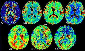
Forty-four patients (33 males and 11 females) diagnosed with brain SVD were recruited as the SVD patient group, twenty-four patients (19 males and 5 females) diagnosed with brain BVD were recruited as the BVD patient group, and 16 age- and education-level matched healthy volunteers (12 males and, 4 females) were recruited as the normalcontrol group. Routine MR scan were performed on a whole body 3T scanner (Ingenia, Philips Healthcare) with a 16-channel dS head coil. Kurtosis images were acquired with the following parameters: TE = 91 ms, TR = 1000 ms, number of slices = 18, maximum b value = 2000 andnumber of directions =32. DKE (Version 2.5.1) was employed to generate kurtosis related parameters (Figure 1). The mean kurtosis (MK), the fractional anisotropy (FA), and the mean diffusion coeficient (MD) of cerebral white matter were compared with t-test between each patient group and the control group in basal ganglia, thalamus, corona radiata, centrum ovale, and the locations beside lateral ventrical, pons and callosum.Results and Discussions
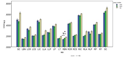
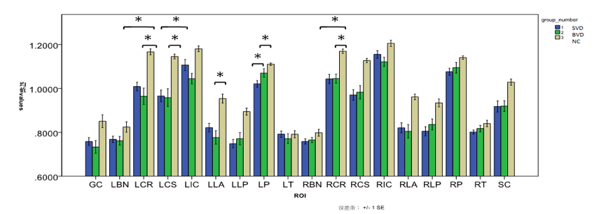
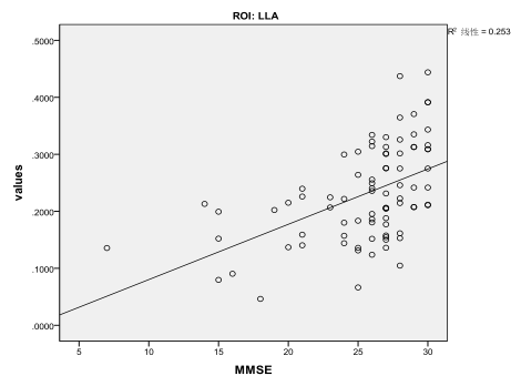
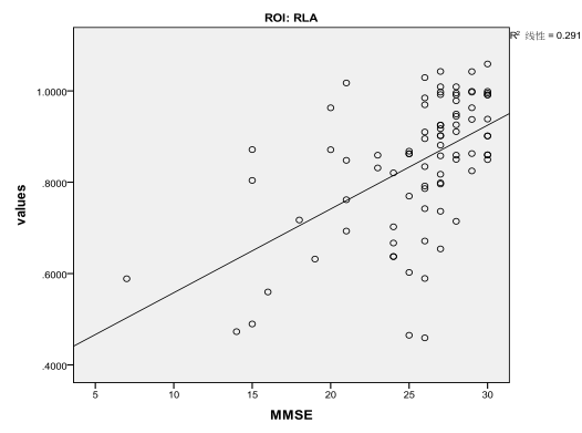
There was significant difference of FA value in the right caudate nucleus between the SVD and BVD groups with the same degree of cognitive impairment (Figure 2). The MD values increased in both SVD and BVD groups, indicating that the infarction reason is angiogenic edema, not cytotoxic edema as before. This is consistent with the pathological change of cerebral ischemia and infarction in the chronic latent progress of SVD and the absence state of large acute arterial occlusion in the ROI. In SVD group, MD values of left thalamus is different from those in BVD (P=0.026), suggesting special diffusion movements of free water molecules in the left thalamus. And parameters of the three groups show consistency. Moreover, MK value decreased in SVD and BVD groups, indicating the structural complexity of the corresponding brain tissue which was damaged in the pathological state. Specially, MK value of left pons in SVD group decreased more obviously than in BVD group (P =0.036) (Figure3). The RK and AK values of the left side of the bridge brain in SVD and BVD groups were dramatically decreased compared to control group, while these values remain closed in the two patient groups. DKI derived parameters and cognitive evaluation scores are linearly correlated (Figure 4, 5). The results suggested that DKI could indicate sensitive developmental changes of local microstructures in brain SVD and BVD. The conventional diffusion parameters were estimated using the mono-exponential model, where the derived values depended on the selection of b values. As an extension of DTI model, DKI required at least two non-zero b values in more than 15 independent directions1. With a second-order polynomial model, DKI could provide a b-value-independent estimation of the diffusion and kurtosis parameters. Therefore, DKI may be an ideal technique for estimating the restricted diffusion process in in vivo study, especially in detecting the pathological alterations in neural tissues2.Conclusion
In conclusion, DKI could provide sensitive developmental changes of local microstructures in brain SVD and BVD. In addition, DKI-derived diffusion parameters are sensitive to alterations in white matter regions with complex fiber arrangements3. The atrophy may exist in white matter fiber. Moreover, DKI can reflect the degree of cognitive impairment caused by cerebral vessel diseases. This is useful in early diagnosis and choice of monitoring strategy, as well as dynamic observation and prognostic assessment.Acknowledgements
This work was supported by a grant from National Natural Science Fund, China (Project No.: 81571231) and Science and technology development project of Jilin Provincial Health Department, China (Project No.: 2015Z043).References
[1] Coutu J P, Chen J J, Rosas H D, et al. Non-Gaussian water diffusion in aging white matter[J]. Neurobiology of Aging, 2014, 35(6):1412-1421.
[2] Zhu J, Zhuo C, Qin W, et al. Performances of diffusion kurtosis imaging and diffusion tensor imaging in detecting white matter abnormality in schizophrenia[J]. Neuroimage Clin, 2015, 7(C):170-176.
[3] Steven A J, Zhuo J, Melhem E R. Diffusion kurtosis imaging: an emerging technique for evaluating the microstructural environment of the brain.[J]. Ajr American Journal of Roentgenology, 2014, 202(1):W26.
Figures