2664
A new accelerated time-of-flight Brain MR angiography (Spiral MRA) with a combination technique of spiral acquisition and fat suppression: ProSet1Department of Diagnostic Imaging and Nuclear Medicine, Tokyo Women's Medical University, Tokyo, Japan, 2Philips Electronics Japan, Ltd., Tokyo, Japan
Synopsis
Spiral MRA is a new accelerated time-of-flight MR angiography (TOF-MRA), the k space is filled with data in a spiral trajectory on the frequency and phase encoding directions. In this study, the effect of TONE and ProSet on Spiral MRA was evaluated by comparing image quality between Spiral MRA and conventional TOF-MRA. As the result, TONE was rarely effective on Spiral MRA, and Spiral MRA with ProSet provided high quality images, and reduced the acquisition time by approximately 70%, compared to conventional TOF-MRA with ProSet. In conclusion, Spiral MRA with ProSet is a useful, accelerated technique without image quality deterioration.
Introduction
Time-of-flight Brain MR angiography (conventional MRA) is commonly combined with TONE to improve demonstration of peripheral arteries.1, 2 In addition, a combination of conventional MRA and fat suppression is not widely used because fat suppression sequences can lead to acquisition time extension and pseudo-stenosis due to longer TR and TE. Spiral MRA is a new time-of-flight MRA based on spiral acquisition in k-space (Figure 1), and is expected to shorten acquisition times while keeping image quality.3 In k-space, MR data is acquired in a spiral trajectory, from the center to the outside on the frequency and phase encoding directions. Therefore, it is suspected that TONE may be less effective on Spiral MRA, compared to conventional MRA, and that a combination of Spiral MRA and the fat suppression sequence ProSet could be useful to improve image quality with minimum acquisition time extension even though ProSet cannot be combined with TONE. In this study, we estimated the effect of TONE and ProSet on Spiral MRA to provide accelerated, high quality MRA.Methods
Five healthy volunteers were examined. (males: 3, females: 2, ages: 30 to 46 years) All studies were performed with a 1.5T MR unit. (Ingenia CX, Philips Healthcare) Image quality of Spiral MRA with TONE, Spiral MRA without TONE, Spiral MRA with ProSet, and conventional MRA with ProSet were evaluated using a 5-point scale and signal profile analysis. The parameters of each MRA are shown in Table 1.Results
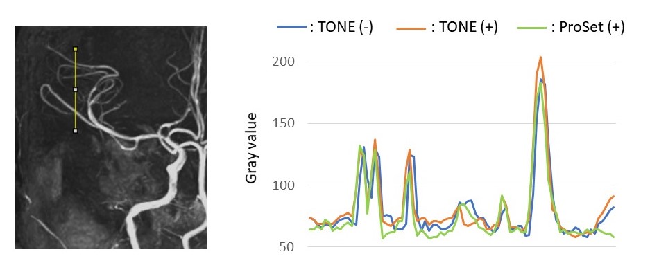
There was no significant difference in image quality between Spiral MRA with TONE and Spiral MRA without TONE. Visualization of external carotid arteries (superficial temporal arteries and occipital arteries), ophthalmic arteries (OphA), and anterior/posterior inferior cerebellar arteries (AICA/PICA) on Spiral MRA with ProSet was significantly improved compared to Spiral MRA without ProSet (with/without TONE) (Figure 2). There was no significant difference in signal profile analysis of middle cerebral arteries (MCA) among each Spiral MRA (Figure 3). Conventional MRA with ProSet (acquisition time: 8min) showed similar image quality as Spiral MRA with ProSet (acquisition time: 3min) (Figure 2).Discussion
Spiral MRA is a new technique, and row MR data is collected into k-space with a spiral trajectory from the center to the outside on both frequency and phase encoding directions. In this study, ProSet was useful to improve demonstration of each artery. Especially, external carotid arteries, OphA, AICA and PICA were clearly noted on Spiral MRA with ProSet. However, signal profile analysis of MCA on each Spiral MRA showed similar results. Therefore, we suggested that TONE rarely affected the demonstration of peripheral arteries on Spiral MRA because TONE is applied within the slab, and that ProSet was very effective for Spiral MRA to improve the image quality because it suppressed high signals from fat on the background. Conventional MRA with ProSet was just as good as Spiral MRA with ProSet. However, conventional MRA with ProSet caused excessive acquisition time extension, and Spiral MRA with ProSet was able to reduce the acquisition time by approximately 70%, compared to conventional MRA with ProSet. Therefore, we suggested that Spiral MRA with ProSet has a big advantage to be a new accelerated MRA.Conclusion
A combination of Spiral MRA and ProSet will contribute as a new accelerated technique providing high quality images.Acknowledgements
No acknowledgement found.References
1. Domoulin CI, Cline HE, Souza SP, et al: Three-dimensional time-of -flight magnetic resonance angiography using spin saturation. Magn Reson Med 11:35-46, 1989
2. Atkins D, et al. Improved MR angiography: Magnetization transfer suppression with variable flip angle excitation and increased resolution. Radiology, 190: 890-894, 1994
3. Fielden SW1, Meyer CH. A simple acquisition strategy to avoid off-resonance blurring in spiral imaging with redundant spiral-in/out k-space trajectories. Magn Reson Med 73:704-10, 2015
Figures


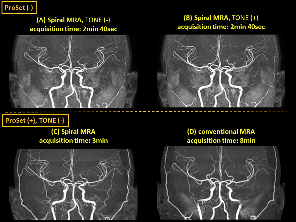
Figure 2.
(A) Spiral MRA, TONE (-), ProSet (-) (B) Spiral MRA, TONE (+), ProSet (-) (C) Spiral MRA, TONE (-), ProSet (+) (D) conventional MRA, TONE (-), ProSet (+)