2663
Optimization of a new accelerated time-of-flight Brain MR angiography using spiral data acquisition: Spiral MRA1Department of Radiological Services, Tokyo Women's Medical University Hospital, Tokyo, Japan, 2Department of Diagnostic Imaging and Nuclear Medicine, Tokyo Women's Medical University, Tokyo, Japan, 3Philips Electronics Japan, Tokyo, Japan
Synopsis
Spiral MRA is a new accelerated time-of-flight MR angiography (TOF MRA) with spiral data acquisition, which acquires MR data by traveling through k-space with spirals. Acquisition window (AW) is a new parameter, which indicates the degree of under-sampling related to image quality and acquisition time. In this study, suitable flip angle (FA) and AW for Spiral MRA was evaluated by a 5-point scale and signal profile analysis. In conclusion, the suitable FA was 25° to demonstrate each artery and suppress the background signals. AW should be set to 10 or less to avoid vessel blurring.
Introduction
Time-of-flight Brain MR angiography (TOF MRA) is a widely used technique,1 however, a new technique is required to shorten the acquisition time. Recently, Spiral MRA has been developed as a new accelerated TOF MRA.2 On Spiral MRA, k-space is filled with MR data in a spiral trajectory from the center to the outside on both frequency and phase encoding directions (Figure 1). It is suspected that the suitable flip angle (FA) on Spiral MRA should be different from conventional TOF MRA. In addition, acquisition window (AW) is a new parameter, which determines the speed of filling k-space. It indicates the degree of under-sampling, which can affect image quality and acquisition time. Therefore, we optimized FA and AW for Spiral MRA to create high quality MRA with shorter acquisition time.Methods
Five healthy volunteers were examined. (males: 3, females: 2, ages: 30 to 46 years) All studies were performed with a 1.5T MR unit. (Ingenia CX, Philips Healthcare) Each Spiral MRA was evaluated by a 5-point scale and signal profile analysis to determine suitable FA and AW.
1. Assessment of optimized FA
Spiral MRA with FA:15, 20, 25, 30, 35 were obtained. The other parameters were as follows: TR/TE=24/4.3 ms, FOV=230x230x99 mm, slice thickness=0.45 mm, slice number=220, acquisition window=8, acquisition time=3min 20sec, ProSet (+)
2. Assessment of optimized AW
The imaging parameters were shown in Table 1. FA was decided by the first assessment.
Results
1. Assessment of optimized FA
Each artery on Spiral MRA with FA: 15, 20, 25 was clearly demonstrated, however, visualization of Spiral MRA with FA: 30, 35 was worse due to spotty signal loss. An increase in the background signals on Spiral MRA with FA: 15, 20 was noted, compared to Spiral MRA with FA: 25, 30, 35. Spiral MRA FA: 25 showed a good balance between signal suppression in the background and demonstration of each artery (Figure 2).
2. Assessment of optimized AW
Demonstration of peripheral arteries on Spiral MRA with AW: 14 was significantly worse than Spiral MRA with AW: 6, 8, 10, 12. Vascular blurring and a decrease in gray value was noted on Spiral MRA with AW: 12, 14 compared to Spiral MRA with AW: 6, 8, 10. There was no significant difference in the signal profile analysis among Spiral MRA with AW: 6, 8, 10 (Figure 3).
Discussion
FA is a very important parameter for TOF MRA because it greatly affects in-flow effects and demonstration of peripheral arteries. As for Spiral MRA, high FA: 30, 35 caused spotty signal loss of each artery. Conversely, low FA: 15, 20 increased in the background signals. Therefore, we suggested that the suitable FA was 25 degrees, which is a slightly higher FA, compared to conventional TOF MRA.
AW is an original parameter for Spiral MRA, and determines the speed for filling k-space, in which MR data is acquired with a spiral trajectory. In this study, excessive high AW caused vascular blurring and low in-flow effects, even though it was possible to shorten acquisition time. Spiral MRA acquires MR data on both frequency and phase encoding directions. The higher the AW is set, the lower the MR data is sampled. Therefore, we suggested that the reason may come from the vascular flow, the data collecting method, and low data sampling, and that the AW should be set to 10 or less (Figure 4).
Conclusion
Spiral MRA with FA: 25 and AW: 10 and under can provide high quality images with shorter acquisition time.Acknowledgements
No acknowledgement found.References
1. Domoulin CI, Cline HE, Souza SP, et al: Three-dimensional time-of -flight magnetic resonance angiography using spin saturation. Magn Reson Med 11:35-46, 1989
2. Fielden SW1, Meyer CH. A simple acquisition strategy to avoid off-resonance blurring in spiral imaging with redundant spiral-in/out k-space trajectories. Magn Reson Med 73:704-10, 2015
Figures
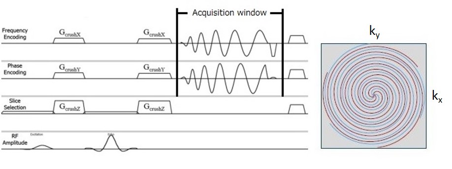
Figure 1. Illustration of Spiral sequence chart
MR data is collected into k-space travelling with a spiral trajectory from the center to the outside on both frequency and phase encoding directions.
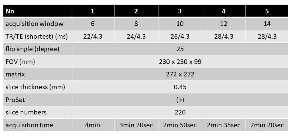
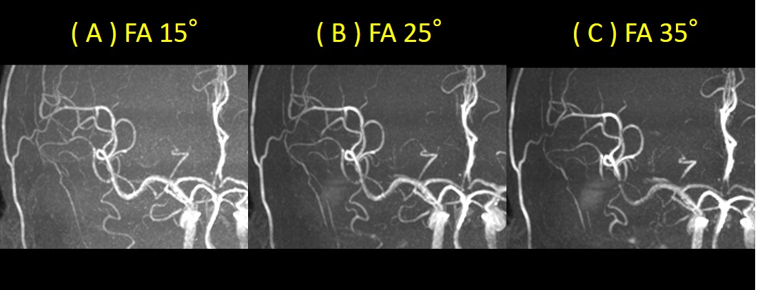
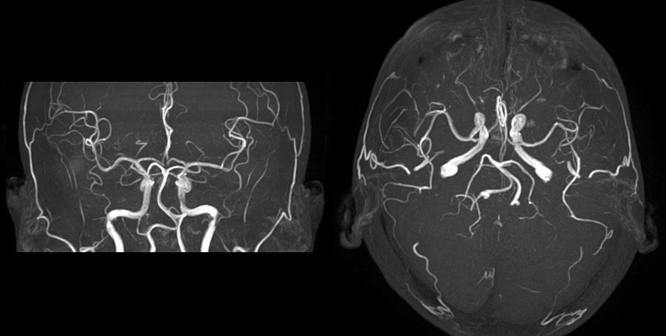
Figure 2. Spiral MRA
Spiral MRA with FA: 15° (A) shows an increase in the background signals. Spotty signal loss of each artery is noted on Spiral MRA with FA: 35° (C). Spiral MRA with FA: 25° (B) shows good signal suppression in the background and demonstration of each artery. FA: flip angle
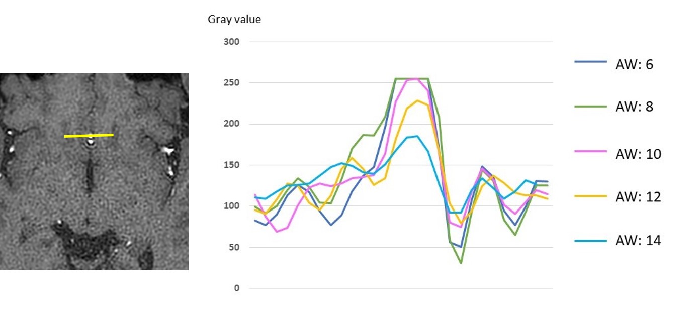
Figure 3. Signal profile analysis of anterior cerebral artery
There is no significant difference in gray values among Spiral MRA with AW: 6, 8, 10. Maximum gray value of Spiral MRA with AW: 12, 14 is lower than Spiral MRA with AW: 6, 8, 10. AW: acquisition window