2655
The value of high-resolution magnetic resonance vascular wall imaging in the diagnosis and treatment of central nervous system vasculitis1Taishan Medical University, Jinan, China, 2Qianfoshan Hospital Affiliated to Shandong University, Jinan, China, 3GE Healthcare, MR Research, Beijing, China
Synopsis
Three-dimensional (3D) CUBE MRI for high-resolution vascular wall imaging can reveal the morphological characteristics of vessel wall. To investigate its feasibility in the diagnosis of central nervous system vasculitis, we applied the contrast-enhanced 3D T1-weighted CUBE imaging in the patients with vasculitis. We found significant alterations of the vessel wall imaging in signal-to-noise-ratio and contrast-to-noise-ratio before and after clinical treatment. With these, we can demonstrate that 3D CUBE MRI can effectively help to diagnose the central nervous system vasculitis and evaluate the treatment effect.
Introduction
Central nervous system vasculitis (CNSV) is an inflammatory lesion that primarily affects the intracranial vascular wall1. So far, digital subtraction angiography (DSA) has been widely used as an imaging method for CNSV diagnosis in the clinic2,3. The sensitivity and specificity of CNSV imaging using DSA is however, not high2,3,4. A three-dimensional (3D) CUBE magnetic resonance imaging (MRI) technique for high-resolution vascular wall imaging has shown its ability to reveal the morphological characteristics of vessel wall5.It remains however, unknown if this technique still has potential in the diagnosis of CNSV. Therefore, this study aimed to explore the feasibility of CUBE MRI in a clinical population confirmed with CNSV. The morphological characteristics of vessel wall as well as the alterations of vessel imaging before and after clinical treatment were investigated systematically.Materials and Methods
Subjects
13 patients (mean age: 50.9 years old, ranged from 27 to73 years old), confirmed with central nervous system vasculitis clinically, were recruited in this study for MRI experiments. Written informed consents were obtained from each patient.
MRI experiment
All experiments were performed on a 3T clinical scanner (Discovery 750w, GE Healthcare, Milwaukee, WI, USA) equipped with a 32channel coil. A contrast-enhanced 3D T1-weighted CUBE technique was employed for vascular wall imaging for each patient before and after clinical treatment. The corresponding scan parameters include:FOV = 200×200mm2;; TR=600ms; TE=14.4ms; slice thickness =1mm; slice gap=0.5mm; NEX=0.5, matrix size=288x288 and echo length train=24. The total scan time was 4 minutes 16s.
Data analysisTo evaluate the alterations of vascular wall imaging before and after treatment, the signal-to-noise ratio (SNR) and contrast-to-noise ratio (CNR) were calculated with the defined equations shown below: SNR= Swall /Snoise [Eq.1]
And, CNR=︱Swall−Slumen︱/SDnoise [Eq.2],
where, Swall, Slumen and Snoise represent the mean signal intensities of the vessel wall, lumen and background, respectively. SDnoise is the standard deviation of the noise.
All statistical analysis were performed in SPSS software 24.0. The embedded McNemar-test was used to evaluate the alterations of the image characteristics before and after treatment, including the signal intensity, ring sign and double track sign. In addition, a paired-sample t test was applied to estimate the difference of the SNR and CNR before and after clinical treatment. The significant threshold was set as p =0.05.
Results
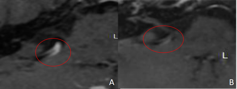
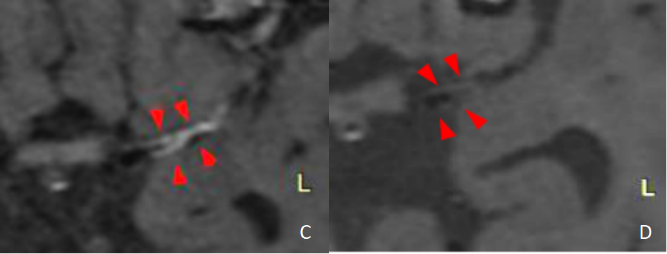
A total of 58 vascular segments involvements were found in 13 patients. The vessel images of patients with CNSV were compared before and after clinical treatment (Figs.1,2). Significantly decreased ring sign and double track sign were found in 47 of 58 patients before treatment. This number was significantly reduced to 16 of 58 after treatment (p<0.001). In addition, the vessel walls with significantly enhanced signal were found in 51 of 58 patients and this number was decreased to 20 of 58 patients after treatment (p<0.001). The SNR and CNR values of vessel wall imaging were also calculated before and after treatment. Significant difference was found before and after treatment(p<0.001).Discussion and conclusion
In this study, contrast enhanced 3D CUBE MRI was applied to image the vessel wall of patients with central nervous system vasculitis before and after clinical treatment. A thickened vessel wall and significantly enhanced signal was found in the patients with vasculitis. Moreover, the ring sign and the double track sign were also found after image enhancement in the patients . In addition, a significantly decreased signal intensity was found in the vessel wall after treatment relative to before treatment, showing significantly reduced patient numbers with ring sign and double track sign. This is agreed with the findings reported by Jennette J et al (2013)6. In addition, the significantly reduced SNR and CNR values in the vessel wall imaging indicate the quantitative alterations of vessel imaging before and after clinical treatment, which also supports the imaging features we have obtained.In conclusion, due to the robust performance on vascular wall imaging before and after treatment shown above, 3D CUBE MRI has been demonstrated to be feasible in the diagnosis of central nervous system vasculitis. It is therefore suggested to be routinely applied as a method in the diagnosis of central nervous system vasculitis.Acknowledgements
No acknowledgement found.References
1.Scolding NJ. Central nervous system vasculitis. Semin Immunopathol 2009; 31: 527–536.
2.Salvarani C, Brown RD Jr, Calamia KT, et al. Angiography-negative primary central nervous system vasculitis: a syndrome involving small cerebral vessels. Medicine (Baltimore) 2008; 87: 264-71.
3.Gomes LJ. The role of imaging in the diagnosis of central nervous system vasculitis. Curr Allergy Asthma Rep 2010; 10: 163–170.
4.Singhal AB, Topcuoglu MA, Fok JW, Kursun O, Nogueira RG, Frosch MP, et al. Reversible cerebral vasoconstriction syndromes and primary angiitis of the central nervous system: clinical, imag-ing, and angiographic comparison. Ann Neurol. 2016;79:882–94.
5.Mandell D M,Mossabasha M,Qiao Y,et al.Intracranial Vessel Wall MRI:Principles and Expert Consensus Recommendations of the American Society of Neuroradiology[J].Ajnr Am J Neuroradiol,2017,38(2):218-229.
6.Jennette JC, Falk RJ, Bacon PA, Basu N, Cid M.C, Ferrario F, et al. 2012 Revised international Chapel Hill consensus conference nomenclature of vasculitides. Arthritis Rheum 2013;65:1-11.
Figures