2649
High resolution MRI in Diagnosis of Cerebral Arterial Thrombosis1Taishan Medical University, Tai'an, China, 2Qianfoshan Hospital Affiliated to Shandong University, Jinan, China, 3MR Research, GE Healthcare, Beijing, China
Synopsis
This study aimed to investigate the feasibility of CUBE MRI for high resolution imaging in the detection of intraluminal thrombi in acute stroke patients. The T1-weighted CUBE images showed dark blood signal in arteries and high signal or iso-signal filling in the lumen. In our study, the sensitivity of T1 weighted CUBE in the detection of intraluminal thrombi reached 100% and the corresponding area under curve(AUC) value was higher than SWI. We therefore demonstrated that the T1-weighted CUBE MRI can effectively help to diagnosis the intraluminal thrombi.
INTRODUCTION
Detecting arterial occlusion has prognostic and therapeutic implications for acute stroke patients1. It is thus, crucial to determine the spatial and morphological information of thrombus in the clinical diagnosis. Magnetic susceptibility weighted (SWI) technique, showing the inherent differences in magnetic sensitivity between tissues, has been reported to show certain significance in the diagnosis of acute cerebral artery thrombosis2.However, Thrombus in many locations cannot be well displayed due to the magnetic susceptibility artifacts and different compositions of thrombi3.
Additionally, CUBE magnetic resonance imaging (MRI), as a three-dimensional (3D) high resolution imaging technique, has been widely applied in the diagnosis of intracranial arterial wall lesions4,5. This technique was thus assumed to be able to diagnose intracranial arterial thrombosis. Therefore, this study aimed to investigate the feasibility of T1-weighted CUBE technique in the detection of intraluminal thrombi in acute stroke patients comparing with SWI images. Digital subtraction angiography (DSA) was also measured and served as a reference.
Materials and Methods
Sixty-nine patients (38 males and 31 females, mean age 53 years old, onset time 8h to 14d) with acute stroke were enrolled in this study. Written informed consents were obtained from each patient. Each patients was evaluated with diffusion weighted imaging (DWI),SWI and T1-weighted CUBE sequences.
All experiments were performed on a 3T clinical scanner (Discovery 750w,GE Healthcare, USA) equipped with a 32-channel coil. 3D CUBE T1 sequence was scanned with scan parameters of TR = 600ms, TE = 14.4ms, thickness = 1mm, gap= 0.5mm, FOV = 200mm * 200mm for whole brain coverage, matrix size = 288 * 288 and echo chain length = 24. Total scanning time was 4 minutes 16s.
DSA was used as the reference standard to establish occlusion of the intracranial artery. Intraluminal thrombus on the T1-weighted CUBE images was defined as the presence of high signal or iso-signal filling in the lumen compared to no signal intensity of the normal vessel. The susceptibility sign on SWI for detecting an intraluminal thrombus was defined as the presence of low signal intensity within the craniocerebral artery, in which the diameter of low signal within the vessel exceeded the contralateral vessel diameter. Two combinations of imaging sequences for detecting an intra-arterial thrombus were set:1.DWI+ T1-weighted CUBE 2.DWI+SWI. The diagnostic results of both sets were compared with the DSA results. The diagnostic value of T1-weighted CUBE and SWI was compared by calculating sensitivity, specificity, positive predictive value, negative predictive value and area under ROC curve (AUC value).
All statistical analyses were performed in MedCalc software. Weighted kappa (κ) statistics was used to assess the agreement between T1-weighted CUBE and DSA. The significant threshold was set as p=0.05.
RESULTS
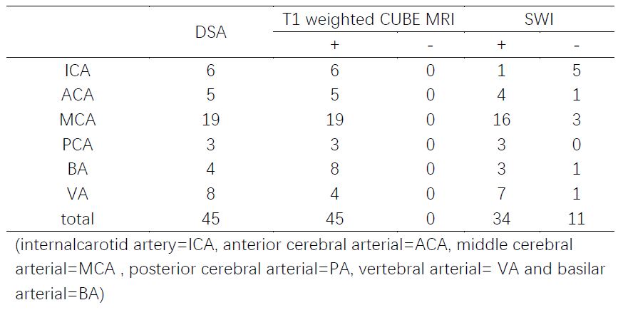
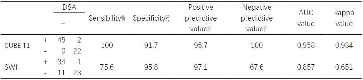
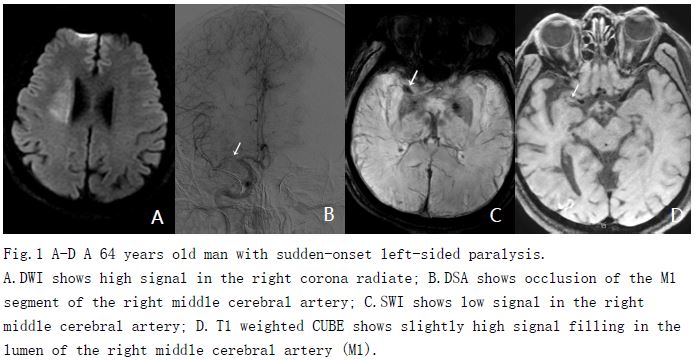
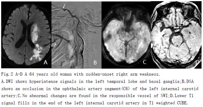
Forty-five patients were diagnosed craniocerebral arterial thrombosis according to DSA examination. All 45 patients were shown positive on T1-weighted CUBE images and only 34 were positive on SWI images (Table 1). In addition, T1-weighted CUBE images showed significantly higher sensitivity (100%vs75.6%, p<0.05) and negative predictive value (100% vs 67.6%, p<0.05) than those of SWI images for detecting intraluminal thrombi (Table 2).The AUC value of T1-weighted CUBE was higher than that of SWI (0.958 vs 0.857, P=0.038, Table 2). T1-weighted CUBE was in a great agreement with the reference standard (Kappa=0.934). Among the patients included in this study, the MCA thrombosis was the most common (19 cases,Table 2), and the diagnostic accuracy of both techniques was high (Fig.1 A-D).For thrombosis in ICA, SWI had the worst diagnostic accuracy (only 1/6 was diagnosed), and T1-weighted CUBE could be all clearly displayed (Fig.2 A-D).DISCUSSION
As a 3D high resolution MRI method, T1-weighted CUBE sequence has been used in this study to visualize the cerebral artery thrombosis. To investigate the feasibility in this application, DSA results, serving as a reference, were acquired and compared with CUBE and SWI images respectively. The diagnostic value of CUBE images were evaluated and compared with SWI. Significantly higher sensitivity and negative predictive value were found in CUBE images and the AUC value of T1 weighted CUBE was much higher than that of SWI. So for the diagnosis of cerebral thrombosis, T1-weighted CUBE is more valuable than SWI. In addition, T1-weighted CUBE was in an excellent agreement with the DSA result, so the feasibility of T1-weighted CUBE images in the diagnosis of cerebral artery thrombosis can be validated.CONCLUSION
In conclusion, T1-weighted CUBE MRI can be demonstrated to be applied for cranial cerebral thrombosis imaging. High clinical value can be validated in the diagnosis of intraluminal thrombi in acute stroke patients.Acknowledgements
No acknowledgement found.References
[1] Derex L, Nighoghossian N, Hermier M, et al. Early detection of cerebral arterial occlusion on magnetic resonance angiography: predictive value of the baseline NIHSS score and impact on neurological outcome[J]. Cerebrovascular Diseases, 2002, 13(4):225-229.
[2] Rovira A,Orellana P,Alvarez-Sabín J,et al.Hyperacute ischemic stroke:middle cerebral artery susceptibility sign at echo-planar gradient-echo MR imaging[J].Radiology,2004,232(2):466-473.
[3]Shinohara Y,Kinoshita T,Kinoshita F,et al.Changes in susceptibility signs on serial T2 ∗-weighted single-shot echo-planar gradientecho images in acute embolic infarction:comparison with recanalization status on 3D time-of-flight magnetic resonance angiography[J].Neuroradiology,2012,54(5):427-434.
[4] Mandell D M,Mossabasha M,Qiao Y,et al.Intracranial Vessel Wall MRI:Principles and Expert Consensus Recommendations of the American Society of Neuroradiology[J].Ajnr Am J Neuroradiol,2017,38(2):218-229.
[5] Li M L,Xu Y Y,Hou B,et al.High-resolution intracranial vessel wall imaging using 3D CUBE T1 weighted sequence[J].European Journal of Radiology,2016,85(4):803-807.
Figures