2646
A high-resolution MRI template for adult Beagle dogXueru Liu1,2, Rui Tian2,3,4, Zhentao Zuo1,2,4, Hui Zhao3,5, Liang Wu2,3, Yan Zhuo1,2,4, Yongqing Zhang2,3,4, and Lin Chen1,2,4
1State Key Laboratory of Brain and Cognitive Science, Institute of Biophysics,Chinese Academy of Sciences, Beijing, China, 2University of Chinese Academy of Sciences, Beijing, China, 3State Key Laboratory of Molecular Developmental Biology, Institute of Genetics and Developmental Biology, Chinese Academy of Sciences, Beijing, China, 4CAS Center for Excellence in Brain Science and Intelligence Technology, Chinese Academy of Sciences, Beijing, China, 5Key Laboratory of Regenerative Biology, South China Institute for Stem Cell, Biology and Regenerative Medicine, Guangzhou Institutes of Biomedicine and Health, Chinese Academy of Sciences, Guangzhou, China
Synopsis
High-resolution T1w and T2w templates from 10 male adult purebred beagles were created in this study. According to the tissue probability map, descriptive statistics of brain tissue volumes and brain sizes exhibit our template with smaller variance. Significant correlation between brain size from dorsal to ventral and gray matter volume was found. This high-resolution purebred canine brain template lays the foundation for further studies aimed at in-vivo analysis of the development of canine brain anatomy and function.
Introduction
Recently, neuroscience research has been extended from non-human primate and rodent models to the domestic dog, who exhibits similar emotion and social processing as human1. Furthermore, the domestic dog is potentially a promising animal model by genetic manipulations for understanding neural mechanism of psychiatry diseases such as autism. MRI/fMRI as one of the powerful tools can non-invasively explore anatomic variation and functional activation. Luckily, several groups published population-averaged canine brain templates2, 3, 4, 5. However, the dogs they used were greatly different in age, skull shape, body size, and brain anatomy, besides there is yet no high-resolution brain template for purebred dogs, which is essential for understanding brain development and function. In this study, high-resolution T1w and T2w brain templates for male adult purebred beagle would be created.Methods
Ten purebred adult beagles (all males, weight 10.54 ± 0.9 kg, age 2.38 ± 0.85 years) were scanned at Siemens prismafit 3.0T MR scanner (Siemens healthnieer, Erlangen, Germany) using home-made 4 channel Tx/Rx coil. Each dog was pre-anesthetized with intramuscular atropine (0.05 mg/kg), followed by Zoletil 50 (10 mg/kg) and Sumianxin (1.6 mg/kg). Complementary anesthesia was performed every one hour with Zoletil 50 (5 mg/kg). T1 weighted, T2 weighted and proton density weighted images in a 3D volume with a field of view of 128 x 128 x 128mm3 and 0.5mm isotropic voxel size were acquired with MPRAGE (magnetization prepared rapid acquisition gradient echoes), T2 space and GRE, respectively. All the experimental protocols were approved by the local institute Animal Care and Committee. Preprocessing was performed using SPM12 (https://www.fil.ion.ucl.ac.uk/spm/software/spm12/), implemented in Matlab (The Mathworks Inc., USA) and FSL (Functional MRI of the brain Software Library). Initially, subjects were re-orientated to AC-PC space and resampled from 0.5 to 0.25 mm isotropic. Then using FSL fast tools to segment T1w image to initial gray matter (GM), white matter (WM), and cerebral spinal fluid (CSF) tissue images, and using SPM to create tissue probability maps (TPM) template. Fig. 1 exhibits the detailed preprocessing steps. In step 4, individual morphological parameters, including GM, WM, and CSF volumes, were extracted based on CAS-Beagle brain template.Results
Fig. 2 demoes a typical beagle dog high-resolution skull-stripped T1w brain image and segmented GM, WM and CSF tissue images in each row. T1w image shows high contrast between GM and WM, clear boundary among GM, WM and CSF. High-resolution purebred beagle T1w and T2w brain templates named as CAS-Beagle Template were created. Fig. 3 shows the sagittal, coronal, and transversal CAS-Beagle TPM overlaid on T1w template, which was generated from 10 male adult beagles, whose brain size are 55.7 ± 2.4mm in the right-left dimension (RL), 81.0 ± 2.6mm in the anterior-posterior dimension (AP) and 45.1 ± 1.8mm in the dorsal-ventral dimension (DV). Multiple-transversal slices of CAS-Beagle TPM overlaid on T1w template are presented in Fig. 4. A 21 parcel atlas5 were normalized to CAS-Beagle TPM and overlaid on template. The descriptive statistics of beagle brain morphology is listed in Fig. 5. GM volume shows significant correlation only with brain size of DV (r2 = 0.7652, p < 0.05, Fig. 5A), the correlation coefficients among other brain morphological parameters are not significant. Only GM volume shows close correlation with body weight (r2 = 0.3067, p < 0.05, Fig. 5B), the correlation coefficients between other brain morphological parameters and body weight are not significant. The absolute volumes of GM, WM, CSF, and WB (whole brain) are 48.10 ± 6.50 mL, 23.47 ± 3.00 mL, 2.48 ± 0.72 mL, and 74.05 ± 8.84 mL, respectively. The mean volumes are very close to that of a previous report, but the standard deviations are smaller than that in the report5. The ratio of GM to WM volume is 2.06 ± 0.28 and GM to WB volume is 0.65 ± 0.002. Tissue volumes (per kg body weight) are 4.60 ± 0.53 mL for GM, 2.25 ± 0.29 mL for WM, 0.24 ± 0.07 mL for CSF, and 7.09 ± 0.76 mL for WB.Discussion & Conclusion
High-resolution purebred beagle MRI T1w and T2w templates were successfully created from 10 adult beagles. This template could potentially provide more insight into the promotion of beagle brain template used in emotional and social processing research on human beings and canines. The CAS-Beagle Template would facilitate future studies to answer important questions, such as the variance of cortical topographies and the longitudinal trajectories of brain development along different ages of different breeds of canines.Acknowledgements
This work was supported in part by the Ministry of Science and Technology of China (2015CB351701), the National Natural Science Foundation of China (31730039, 81871350), National Major Scientific Instruments and Equipment Development Project (ZDYZ2015-2) and Chinese Academy of Sciences Strategic Priority Research Program B grants (XDBS01000000).References
- Bunford N, Andics A, Kis A, et al. Canis familiaris as a model for non-invasive comparative neuroscience. Trends in NeuroScience. 2017; 7(40): 438-452.
- Datta R, Lee J, Duda J, Avants B.B, Vite C.H, Tseng B, et al. A digital atlas of the dog brain. PLoS One. 2012; 7(12): e52140.
- Su M.Y, Tapp P.D, Vu L, Chen Y.F, Chu Y, Muggenburg B, Chiou, J.Y, et al. A longitudinal study of brain morphometrics using serial magnetic resonance imaging analysis in a canine model of aging. Prog Neuropsychopharmacol Biol Psychiatry. 2005; 29(3): 389-397.
- Milne M.E, Steward C, Firestone S.M, Long S.N, O'Brien T.J, Moffat B.A. Development of representative magnetic resonance imaging-based atlases of the canine brain and evaluation of three methods for atlas-based segmentation. American Journal of Veterinary Research. 2016; 77(4): 395-403.
- Nitzsche B, Boltze J, Ludewig E, Flegel T, Schmidt M.J, Seeger J, Barthel H, Brooks O.W, et al. A stereotaxic breed-averaged, symmetric T2w canine brain atlas including detailed morphological and volumetrical data sets. Neuroimage. 2018; 1-11.
Figures
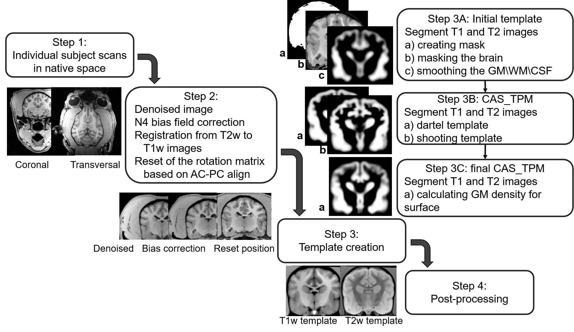
Fig.1. CAS-Beagle Template
processing workflow chart. Step. 1 T1w and T2w MRI brain images were collected
from 10 male adult beagles. Step. 2 All images were reoriented to AC-PC space
after denoised and bias field correction. Step. 3A Segmenting each subject into
GM, WM and CSF and smoothing each tissue. The average smoothed
GM, WM and CSF served as initial TPM. Step. 3B Creating dartel and shooting
template based on Step3A. Step. 3C GM density of brain was calculated for
surface as preparation for post-processing Step (4). AC: Anterior Commissure, PC: Posterior Commissure; GM: Gray
matter; WM: White matter; CSF: Cerebral spinal fluid.
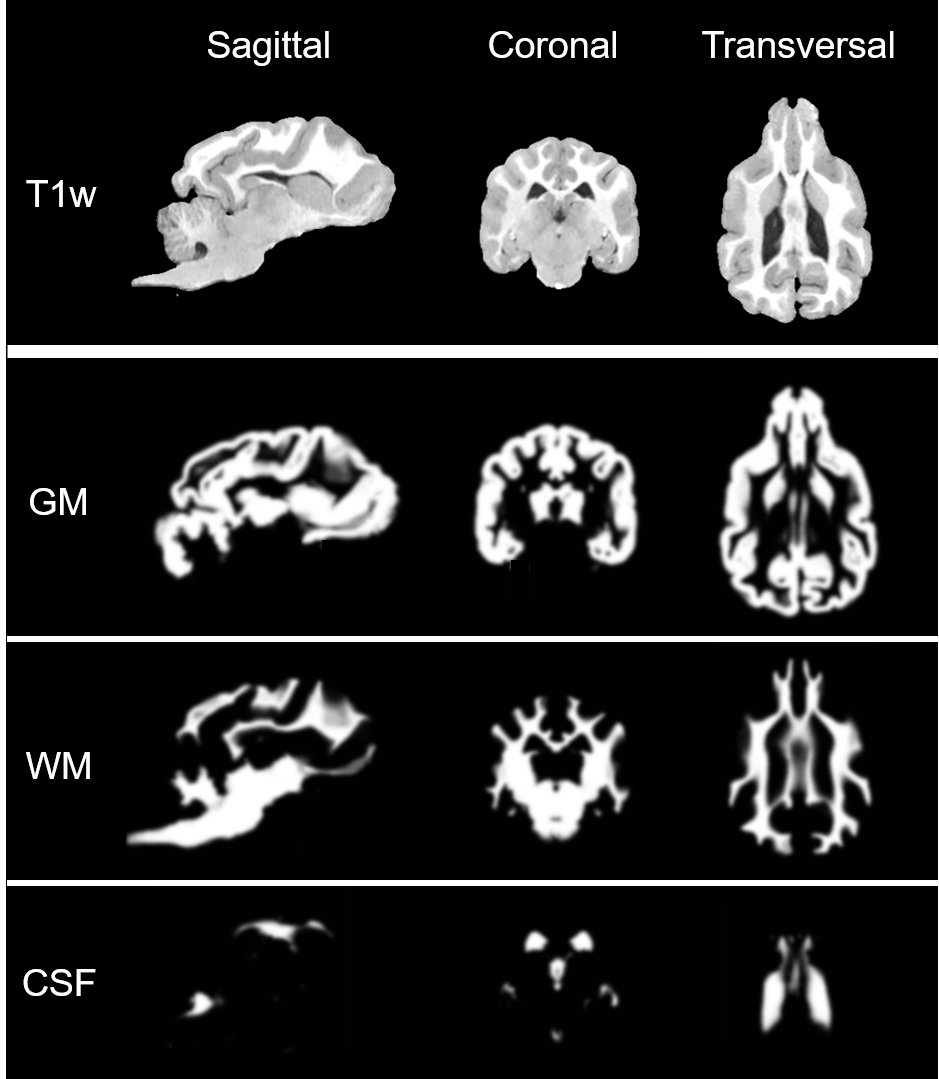
Fig.2. A typical beagle dog T1 weighted original and
segmented tissue map. The upper row presented the original T1 weighted image in
sagittal (left column), coronal (middle column) and transversal (right column).
The corresponding gray matter, white matter and CSF segmented tissue images were
shown on the second, third and fourth rows respectively.
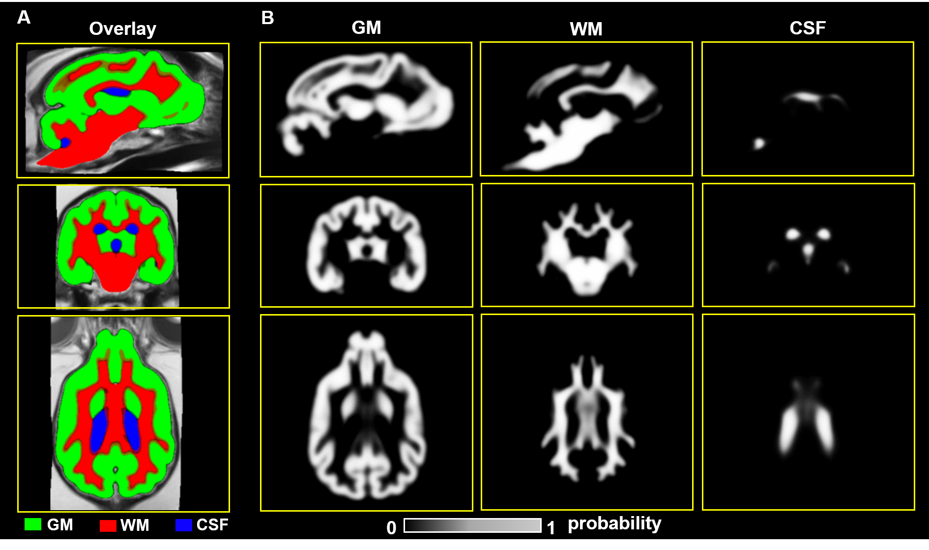
Fig.3. CAS-Beagle Template tissue map. Sagittal,
coronal and transversal sections of the gray matter, white matter and CSF
tissue probability maps were generated from 10 male adult beagles. GM (green),
WM (red) and CSF (blue) are on the second, third and fourth columns,
respectively.
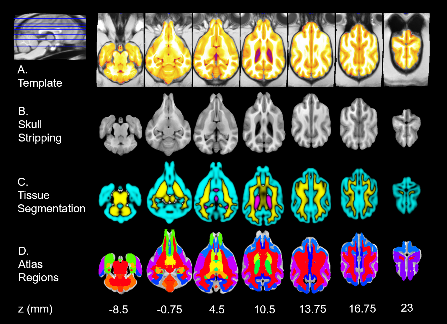
Fig.4. Transversal slices show CAS-Beagle Template.
A) Whole beagle brain template after skull
stripped overlaid on the T1w template. B) The skull stripping beagle brain
template. C) Segmented tissue classes-gray matter (GM; cyan), white matter (WM;
yellow) and cerebrospinal fluid (CSF; violet). D) A 21 parcel atlas5
overlaid on the CAS-Beagle Template.

Fig.5.
Description statistics of the beagle dog brain morphological parameter. (A) GM
volume shows significant correlation with the brain size from dorsal to ventral
(DV). (B) GM volume shows close correlation with body weight. (C) Descriptive
statistics of brain tissue volumes of GM, WM, CSF and WB, and the ratio of GM
volume to WM volume and GM to WB. (D) Descriptive statistics of volume of GM,
WM, CSF and WB per kg body weight.