2645
High-Resolution Magnetic Resonance Imaging of Juvenile Minke Whale Brain at 7T1Translational & Molecular Imaging Institute, Icahn School of Medicine at Mount Sinai, New York, NY, United States, 2Neuroscience, Icahn School of Medicine at Mount Sinai, New York, NY, United States, 3Radiology, Icahn School of Medicine at Mount Sinai, New York, NY, United States
Synopsis
The brain of a 15-foot juvenile brain that washed ashore in the Bronx, New York City was scanned using a 7T whole-body MRI scanner. After fixation in PBS solution and vacuum removal of air pockets, the specimen was scanned using a battery of high-resolution anatomical MRI sequences, including T1-weighted MP2RAGE, 3D MERGE, Proton Density weighted imaging, T2-weighted FLAIR imaging and Diffusion Tensor Imaging. Post-processing included brain masking to alleviate bright background from the PBS solution in most imaging modalities. Anatomical imaging from three high resolution datasets is presented along with a 3D reconstruction generated by volumetric projection of data segmented using the FreeSurfer 6 algorithm.
Introduction
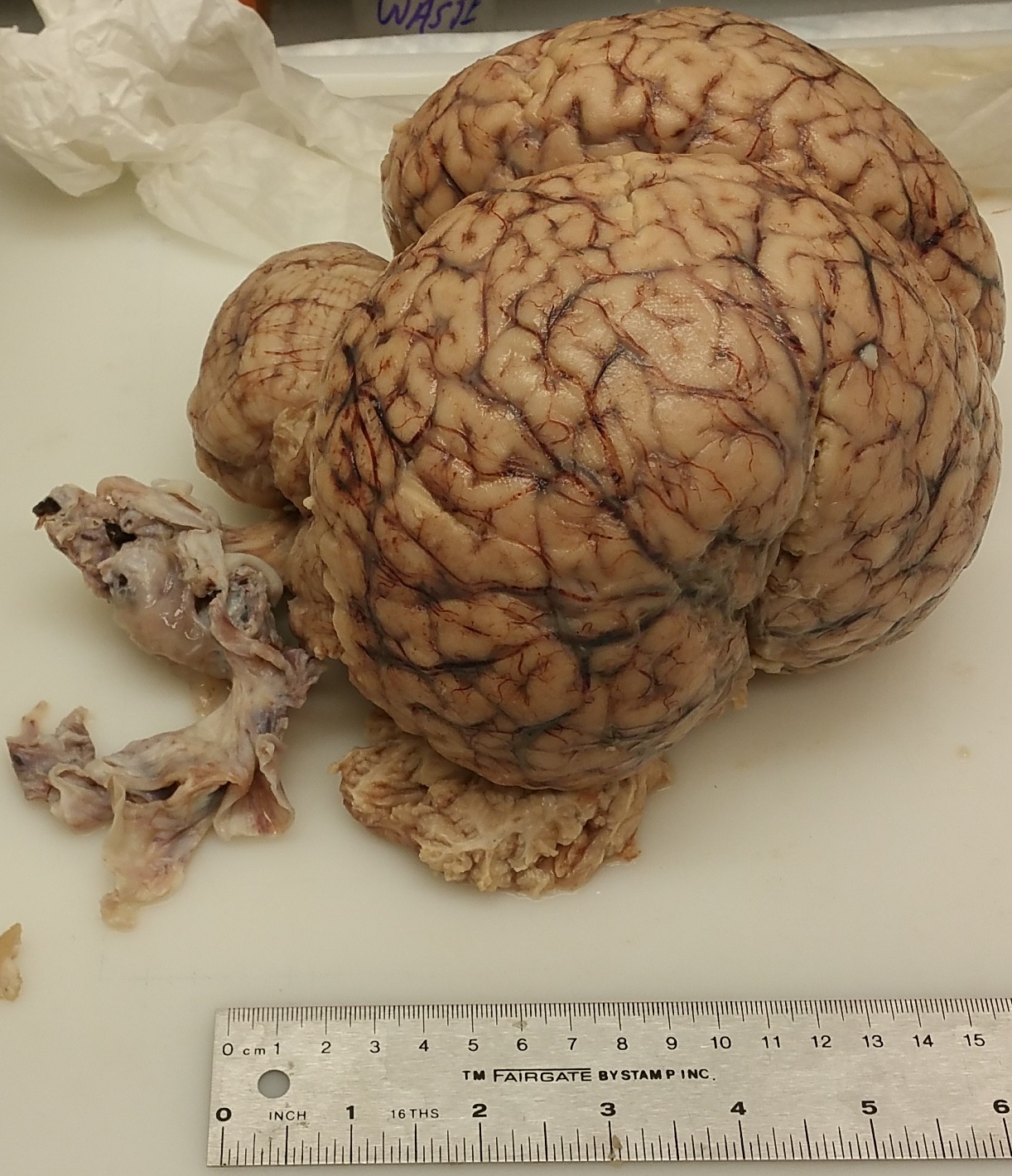
On April 23, 2017, a 15-foot juvenile minke whale washed ashore at Orchard Beach in the Bronx, New York City. While the injured animal was sadly euthanized shortly afterwards, its brain was extracted nearly whole for research through imaging and histology. Figure 1 shows an image of this extracted and preserved brain. Whale brains grow to be much bigger, volumetrically, than human brains, and the neocortex of the minke whale has been found to be similar to that of humans (2.63 mm). However, the layered structure of the whale neocortex is thought to be less intricate, with fewer overall neurons (12.8 billion in minke whales v.s. 20 billion in humans). The total number of neurons in the adult minke whale is approximately 2/3 that of humans, and there are 7.7 times more neocortical glial cells to neocortical neurons in minke whales, compared to a ratio of 1.4 to 1 in humans [1]. There is a large portion in the posterior of the whale brain that has no analogue in human brains. This study describes a unique opportunity to study the developing cetacean brain using high-resolution magnetic resonance imaging at 7T.Materials & Materials
The whale brain specimen measured 17 cm long, 19 cm wide and 12 cm in depth, and was placed in a Phosphate-buffered saline (PBS) solution. A vacuum pump was used to remove pockets of air within the brain to reduce the potential for susceptibility artifacts. The brain was placed in a triple-sealed plastic container with outer labels to serve as imaging landmarks and 2 grams of sodium azide was added to the solution. The brain was then placed inside a 32-channel receive Nova head coil such that its shortest axis (superior-inferior) was aligned along the Z-dimension of the scanner. The brain was braced with padding and tape and scanned over the course of two days.
Scan parameters included:
T1-weighted Magnetization-Prepared 2 Rapid Gradient Echo (T1-MP2RAGE) [2]: TE/TR = 3.98/4000 ms, Field-of-view (FOV) = 200x200 mm, 970x970 matrix size, 240 slices, 0.21 x 0.21 x 0.70 mm voxel size, scan time = 4 hours, 40 minutes.
3D Motion Sensitized Driven Equilibrium prepared Rapid Gradient Echo (MERGE): TE/TR: 4.96/600 ms, FOV = 180x180 mm, 496x496 matrix size, 256 slices, 0.36x0.36x0.40 mm voxel size, 8 averages, scan time = 4 hours, 47 minutes.
Proton Density (PD): TE/TR = 21/10000ms, FOV = 180x180 mm, 960x960 matrix size, 25 slices, 0.19 x 0.19 x 3.0 mm voxel size, 16 averages, scan time = 8 hours, 32 minutes
T2-weighted Fluid Attenuated Inversion Recovery (T2-FLAIR): TE/TR = 117/11010 ms, FOV = 200x200 mm, 35 slices, 1024x1024 matrix size, size, 0.2 x 0.2 x 2.0 mm voxel size, 6 averages, 36 minute scan time.
Diffusion Weighted Imaging: TE/TR = 76.4/15000 ms, B-value = 1500 s/mm2, 68 directions, 69 slices, 0.9 x 0.9 x 1.0 mm voxel size, 10 minute scan time.
A T2-weighted turbo spin echo (TSE) with analagous scan parameters to the T2-FLAIR was also performed though data acquired from the study proved unviable to due to the extremely high background signal from the PBS solution. Brain masks were manually delineated to facilitate removal of the bright background signal due to the PBS present in most of the imaging modalities. Automatic segmentation of gray and white matter was also performed using the FreeSurfer 6 algorithm, although the performance of the algorithm, may not be reliable as it is optimized for human brain segmentation.
Results
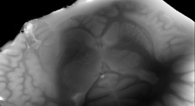
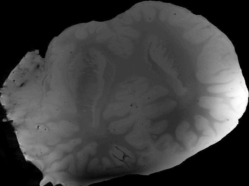
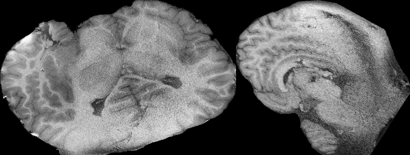
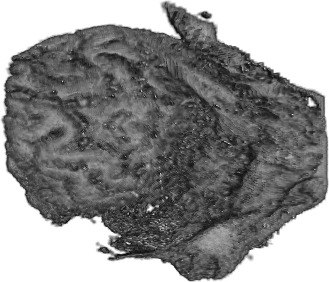
Figure 2 shows an axial slice of a proton-density weighted image with in-plane resolution of around 0.2 x 0.2 mm. Figure 3 shows an axial slice of 3D MERGE data near the level of the ventricles with in plane resolution of 0.36 x 0.36 mm. Figure 4 shows uniform density (UNI-DEN) reconstruction of the MP2RAGE data with in-plane resolution of 0.21 x 0.21 mm. Figure 5 shows the results of Freesurfer segmentation of T1-weighted MRI of the whale brain presented through volumetric projections using Matlab-based macros. The algorithm was still able to identify major cortical structures despite the absence of a skull and presence of bright PBS background.Conclusions
The whale brain posed multiple challenges that provided some insight into the difficulties MR scanning. As a second example, landmarking proved critical in the case of dramatic anatomical abnormalities. Small air pockets resulted in suceptibility artifacts visible across some of the imaging studies, and the brain’s large size renforced the challenges of B1 inhomogeneity for large excitation volumes at ultrahigh field. However, the T1-weighted images produced visualizations of the highly folded cortex of the Minke whale brain in exquisit resolution, as seen in Figure 2.Acknowledgements
Icahn School of Medicine Capital Campaign Translational and Molecular Imaging Institute.References
1. Eriksen, N. and B. Pakkenberg, Total neocortical cell number in the mysticete brain. The Anatomical Record: Advances in Integrative Anatomy and Evolutionary Biology: Advances in Integrative Anatomy and Evolutionary Biology, 2007. 290(1): p. 83-95.
2. Marques, J.P., et al., MP2RAGE, a self bias-field corrected sequence for improved segmentation and T1-mapping at high field. Neuroimage, 2010. 49(2): p. 1271-1281.
Figures