2640
An anatomical atlas for segmentation of thalamic nuclei from conventional 3T MRI1Medical Imaging, University of Arizona, Tucson, AZ, United States, 2Neurology, University of Arizona, Tucson, AZ, United States
Synopsis
Thalamic nuclei are typically invisible on conventional T1 and T2 MRI. We propose here an anatomical atlas based on 7T White matter nulled MP-RAGE data which can be used for a variety of applications including targeting the VIM nucleus for neurosurgical applications and thalamic nuclear volumetry for tracking disease progression using conventional MRI sequences like MP-RAGE or FLAIR.
Introduction
The thalamus and thalamic nuclei play a central role in regulation of consciousness and sleep as well as serve as a critical "switchboard", relaying signals to the cerebral cortex. Various neurological and neuropsychiatric disorders like Parkinson's, schizophrenia, and essential tremor have been atribThe thalamic nuclei are mostly invisible on conventional T1 and T2 MRI sequences like MP-RAGE and fast spin echo (FSE). We adapted the atlas based thalamic segmentation method developed for white matter nulled (WMn) MP-RAGE images for segmenting conventional MP-RAGE images as well as 2D FLAIR images acquired at 3T.Methods
Proposed segmentation method: We generalized the multi-atlas label fusion based THOMAS method previously demonstrated1 for WMn MP-RAGE imaging. The multi-atlas comprises of 20 7T WMn MP-RAGE datasets segmented manually by a neuroradiologist using the Morel atlas as a guide2. A mean template was created by registering and averaging the 20 datasets. The input image was registered to each of the 20 priors via the template to reduce the number of nonlinear registrations. The 20 sets of atlas labels were then warped to the input and combined using majority voting. As an alternative, the mean template was segmented using ST THOMAS and the single set of labels warped to the input after nonlinear registration. While multi-atlas is more robust, the template had very high SNR. We tested it robustness by comparing it with the multi-atlas method.
Segmentation based on conventional MP-RAGE: 18 healthy subjects were scanned on a 3T Siemens Skyra scanner using conventional and WMn MP-RAGE after prior informed consent. The WMn based segmentation was considered the ground truth and compared with the 2 different segmentations (multi-atlas and template) produced from conventional MP-RAGE.
Application to FLAIR imaging: Patients imaged after acute stroke and showing clinical symptoms of amnesia were retrospectively analyzed to assess the extent and nature of thalamic involvement. Thalamic nuclei were segmented from the FLAIR images after nonlinear registration to the mean template and warping the template labels as described above.
Results
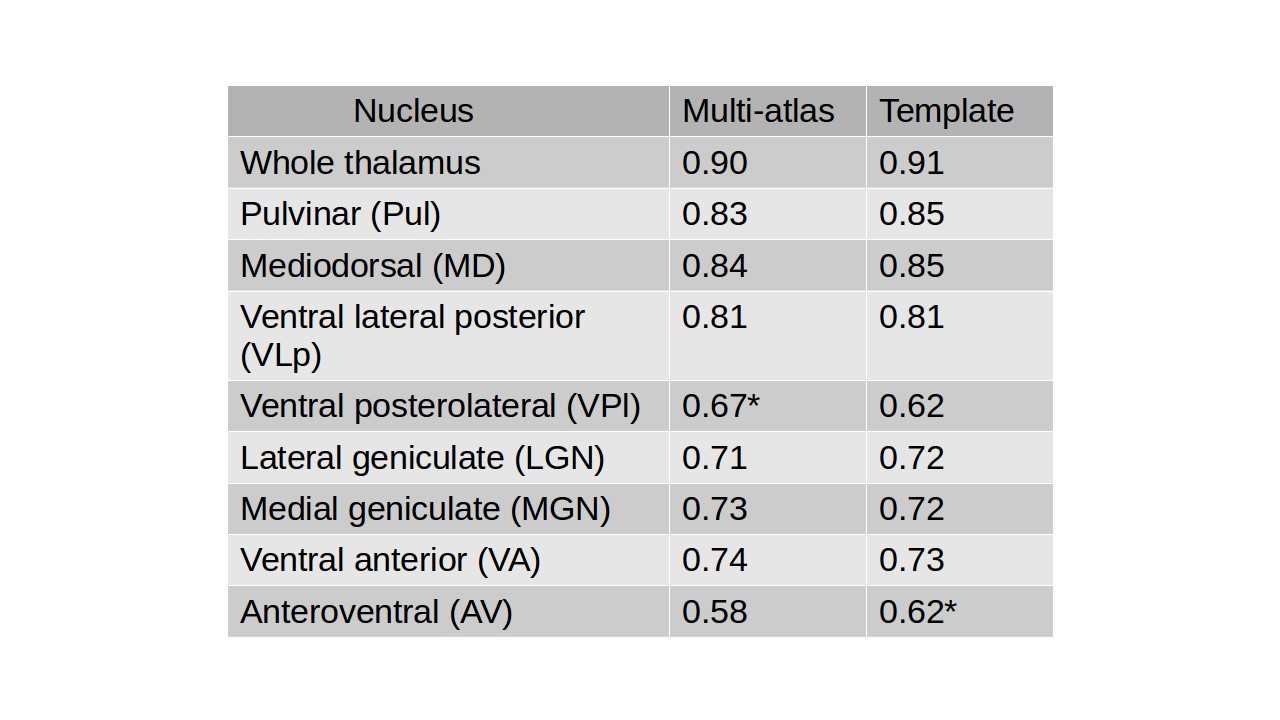
Table 1 shows a comparison of Dice coefficients for thalamic nuclei segmented from conventional MPRAGE using the WMn MPRAGE segmented labels as ground truth. It can be seen that Dice of >0.8 was achieved for larger nuclei such as pulvinar, mediodorsal and ventral lateral posterior. Even for small nuclei such as LGN and MGN, Dice of 0.7 or higher was achieved attesting to the accuracy of the segmentation. There were no significant differences between the multi-atlas and template based labels except for VPl and AV.
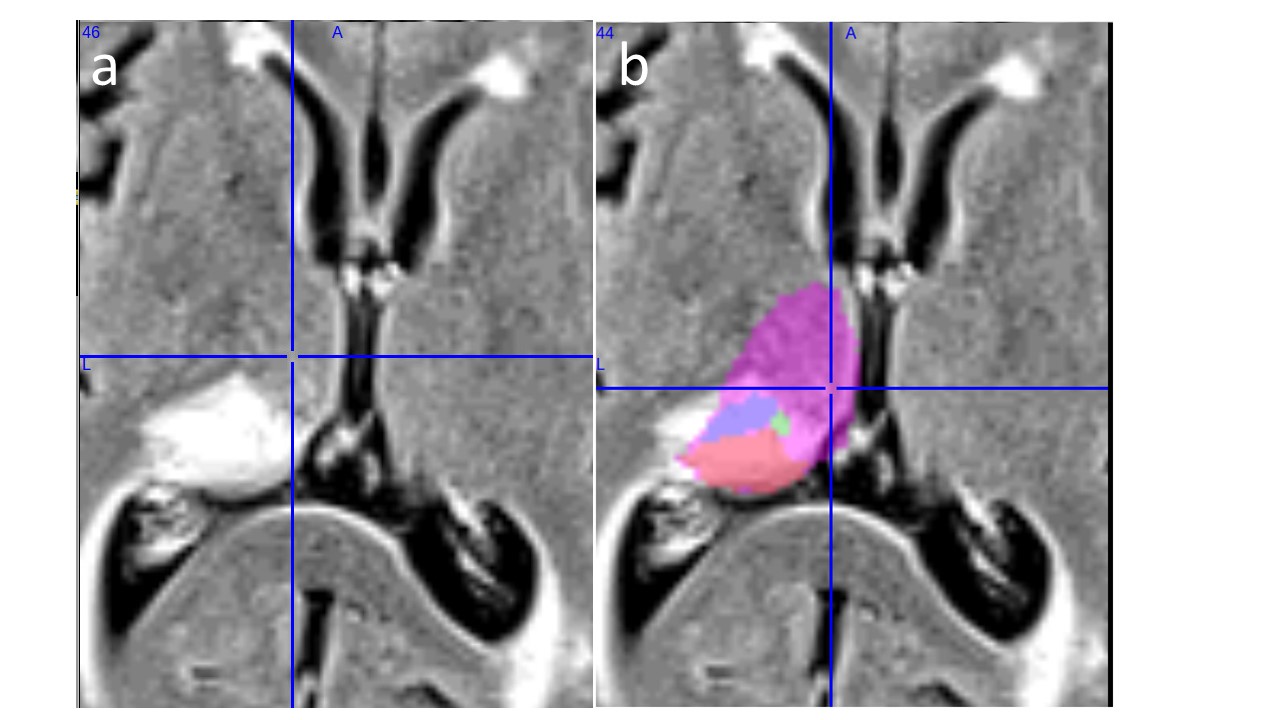
Figure 2a shows FLAIR images from a 79 yr old patient who exhibited sudden confusion and memory impairment showing posterior thalamus involvement. Neurological examination was significant for severe anterograde and retrograde amnesia and right upper quadrantanopia. Figure 2b shows the overlaid thalamic labels (Pul-red, VPL-blue, Centromedial-green, whole thalamus-pink) showing clear non--involvement of anterior thalamic nuclei or mammillothalamic tract. The memory loss was attributed to bilateral medial temporal lobe damage.
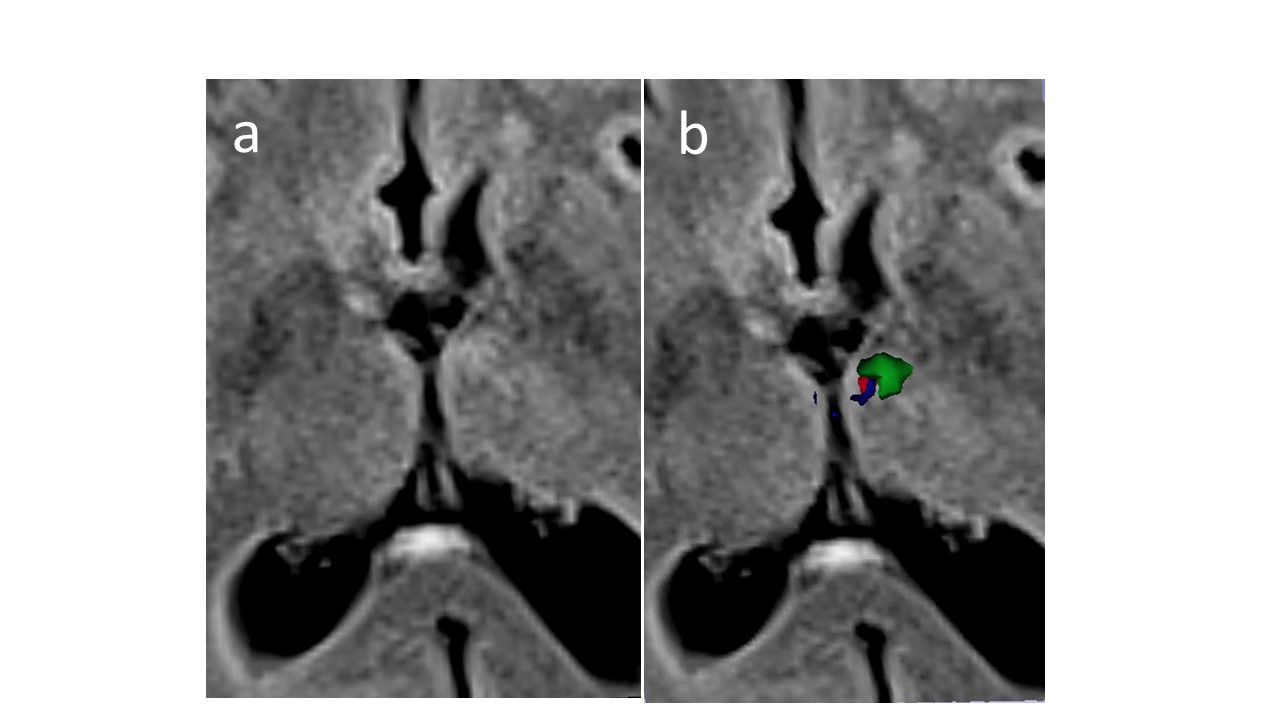
Figure 3a shows FLAIR images from a 70 yr old patient who presented with sudden onset of speech impairment and memory loss. Neurological evaluation demonstrated anomia and severe anterograde and retrograde memory impairment. Figure 3b shows the overlaid thalamic labels (Anterior ventral-red, Ventral anterior-green, MTT-blue) showing clear thalamic involvement of anterior nuclei and MTT in the memory loss.
Discussion and conclusions
This is the first work to use conventional MRI data like MP-RAGE and FLAIR for thalamic nuclei segmentation using an anatomic atlas that was developed using specialized WMn MPRAGE at 7T. Excellent concordance between thalamic segmentation from conventional and WMn MP-RAGE was achieved as shown by the high Dice coefficients. This study has important implications- the WMn MP-RAGE sequence is not widely available on commercial MRI scanners and this and thalamic segmentation using conventional MP-RAGE or even FLAIR is now possible. Furthermore, MP-RAGE data from public databases such as ADNI can be mined for thalamic nuclear volumetry to identify more sensitive biomarkers for diseases such as schizophrenia and Alzheimer's compared to whole thalamic volumes.
Acknowledgements
NIH grant R21 AA023582References
1. Thomas FT, Su J, Rutt BK, Saranathan M. A method for near realtime automated segmentation of thalamic nuclei. Proc. of the 25th scientific meeting of the ISMRMl Hawaii 2017, p4736
2. Tourdias T, Saranathan M, Levesque IR, Su J, Rutt BK. Visualization of intra-thalamic nuclei with optimized white-matter-nulled MPRAGE at 7T. NeuroImage. 2014;84:534-45.
Figures