2638
Visualizing and Characterizing the Habenula with Magnetic Resonance Imaging1Department of Radiology, Ruijin Hospital, Shanghai Jiao Tong University School of Medicine, shanghai, China, 2Magnetic Resonance Innovations, Inc., Bingham Farms, MI, United States, 3Department of Functional Neurosurgery, Ruijin Hospital, Shanghai Jiao Tong University School of Medicine, Shanghai, China, 4Department of Radiology, Wayne State University, Detroit, MI, United States
Synopsis
The habenulae are a small pair of nuclei which serve as a hub between the limbic forebrain and midbrain monoameric neurons. It is a target for the treatment of major depressive disorder using deep brain stimulation, which requires precise pre-treatment mapping. We visualized and characterized the habenula using multiple MRI contrasts and maps to quantify its properties and delineate the structure between lateral and medial side. Axially, we observed elevated iron in the posterior aspect, which we believe to be the lateral habenula. Quantitatively, we also noted similarities of the lateral habenula specifically to white matter.
INTRODUCTION
The habenulae (Hb) consist of a small pair (~30 mm³ per side) of nuclei which bridge the limbic forebrain and midbrain monoamineric centers. They are implicated in major depressive disorder due to abnormal phasic response when provoked by a conditioned stimulus1. The lateral aspect is believed to be involved in dopamine metabolism and is now a target for deep brain stimulation, a treatment which has shown promising anti-depression effects. Prior to DBS treatment, the Hb have to be mapped out in vivo precisely for electrode placement2. Thus, sufficient contrast is of critical importance in both locating it and differentiating lateral from medial components. When imaging subcortical structures, it is recommended that a multi-modal imaging protocol be used to generate different tissue contrasts3. The purpose of this work is to visualize and characterize the Hb using a multi-modal MRI protocol with multiple contrasts and parametric maps to quantify its tissue properties, and to delineate the structure into lateral and medial regions. Having this additional characterization may prove useful in mapping the structure for surgical planning.METHODS
This study was IRB-approved and all subjects were healthy and signed consent forms. All volunteers were imaged with 3 Tesla. Using repeat T1-weighted scans, multi-echo susceptibility weighted imaging (SWI), and strategically acquired gradient echo (STAGE) imaging4,5, we obtained high-resolution, high-SNR T1-weighted imaging, T1 maps, proton spin density (PSD) maps, R2* maps, and quantitative susceptibility maps (QSM)6. ROIs were traced on the T1W images, T2* magnitude, QSM and R2* maps in which volumes from T1W and T2* magnitude images and mean susceptibility and R2* measurements were calculated. A signal threshold was applied to the R2* and QSM images to exclude noise and calculate means. For the T1 and PSD maps, line profiles through the Hb and surrounding gray and white matter tissue and fornix were used to get mean values to account for partial volume effects from CSF and the thalamus.RESULTS
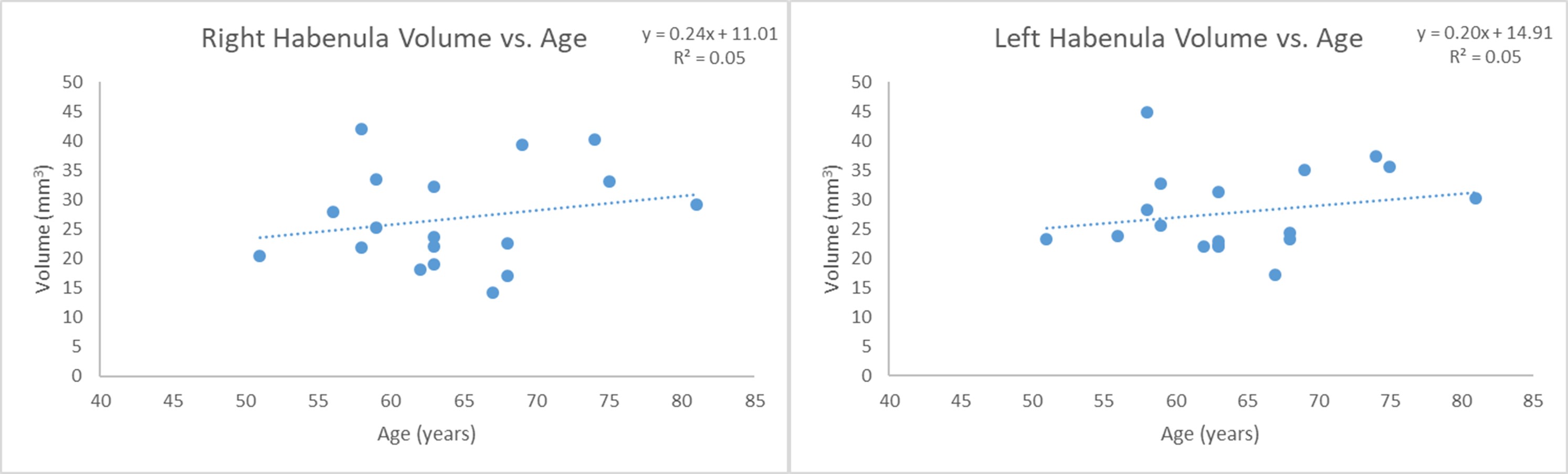
Data from 10 high-resolution T1W scans were averaged to create a high-SNR image set. On these images, we were able to delineate the Hb clearly in the axial view with the medial edge showing a high-contrast appearance as it appears in the triangular depression of the 3rd ventricle; the lateral edge having an arced appearance (Fig. 1). QSM, and R2* showed signal changes in the posterior location indicating high putative iron content within that region (Fig. 2.) Visually, the Hb appeared with similar signal contrast as the fornix in PSD maps and T1 maps (Fig. 2). T2* imaging including the QSM showed small veins which course through and around the Hb. SWI magnitude images showed volumes of roughly 27 mm³ per side. SWI (Fig. 3). Calcification of the pineal gland, located below the Hb, was observed and manifested as blooming and aliasing effect on SWI and QSM.DISCUSSION
The main finding in this work is the location of the increased susceptibility and R2* values in the posterior aspect of the Hb suggest increased putative iron content within the lateral Hb. While this could be suggestive of demyelination, it may also be due to normal aging, give the older age of the volunteers. Anatomically, this study is the first to delineate the structure axially rather than coronally at 3T. The findings from our volumetric analysis were consistent with current literature1,7,8. In T1 and PSD maps, the Hb appeared with similar signal contrast as the fornix suggesting it is predominantly white matter in nature. Venous signal and pineal gland calcification may be confounding to the Hb contrast when viewed with SWI.CONCLUSION
Using 3T MRI, it is possible to map the Hb to delineate the whole structure with T1W along with lateral and medial components, using SWI and QSM maps. This may assist clinicians with pre-treatment mapping of the structure for guided surgery. Further, the presence of veins within and surrounding the structure may be confounding factors in previous fMRI studies which have found anomalous or disrupted responses of the Hb in depression and bipolar disorder.Acknowledgements
No acknowledgement found.References
1. Lawson RP, Nord CL, Seymour B, et al. Disrupted habenula function in major depression. Mol Psychiatry 2017; 22(2): 202-8.
2. Schneider TM, Beynon C, Sartorius A, Unterberg AW, Kiening KL. Deep brain stimulation of the lateral habenular complex in treatment-resistant depression: traps and pitfalls of trajectory choice. Neurosurgery 2013; 72(2 Suppl Operative): ons184-93; discussion ons93.
3. Forstmann BU, Keuken MC, Schafer A, Bazin PL, Alkemade A, Turner R. Multi-modal ultra-high resolution structural 7-Tesla MRI data repository. Sci Data 2014; 1: 140050.
4. Chen Y, Liu S, Wang Y, Kang Y, Haacke EM. STrategically Acquired Gradient Echo (STAGE) imaging, part I: Creating enhanced T1 contrast and standardized susceptibility weighted imaging and quantitative susceptibility mapping. Magn Reson Imaging 2018; 46: 130-9.
5. Wang Y, Chen Y, Wu D, et al. STrategically Acquired Gradient Echo (STAGE) imaging, part II: Correcting for RF inhomogeneities in estimating T1 and proton density. Magn Reson Imaging 2018; 46: 140-50.
6. Li W, Wu B, Liu C. Quantitative susceptibility mapping of human brain reflects spatial variation in tissue composition. Neuroimage 2011; 55(4): 1645-56.
7. Lawson RP, Drevets WC, Roiser JP. Defining the habenula in human neuroimaging studies. Neuroimage 2013; 64: 722-7.
8. Savitz JB, Nugent AC, Bogers W, et al. Habenula volume in bipolar disorder and major depressive disorder: a high-resolution magnetic resonance imaging study. Biol Psychiatry 2011; 69(4): 336-43.
9. Naidich TP, Duvernoy HM. Duvernoy's atlas of the human brain stem and cerebellum : high-field MRI : surface anatomy, internal structure, vascularization and 3D sectional anatomy. Wien ; New York: Springer; 2009.
Figures