2631
Evaluating lifespan tissue structure: Comparing CSD signal fraction and VBM grey matter density1Radiology and Medical Imaging, University of Virginia, Charlottesville, VA, United States, 2University of Virginia, Charlottesville, VA, United States
Synopsis
Constrained spherical deconvolution (CSD), a recently developed diffusion MRI analysis technique, can be used to obtain whole-brain signal fractions from grey-matter-like, white-matter-like, and CSF-like tissue. This study evaluates the CSF compartment present in grey matter (GM-CSF) over the lifespan, and compares it to grey matter density (GMD), obtained through Voxel Based Morphometry. Results of this study reveal a complimentary relationship between GM-CSF and GMD across the lifespan, but not amongst a younger cohort. Results suggest further research is necessary to understand differences between these techniques, and how they may relate to tissue structure.
Introduction
Constrained spherical deconvolution (CSD)-based signal fraction analysis has previously been utilized as an effective means of removing signal in diffusion MRI (dMRI) data that arises from compartments of isotropically diffusing water in grey matter (GM) or cerebrospinal fluid (CSF), in order to better elucidate white matter (WM) signal and directionality1. Recently, using an unsupervised algorithm to determine response functions directly from the dMRI data2, a quantitative utilization of 3-tissue CSD to estimate the WM/GM/CSF signal fractions was demonstrated in white matter hyperintensities in Alzheimer’s disease3,4. CSD is thought to sensitively measure changes in tissue microstructure, but it is not known how the CSD-derived CSF signal fraction compares to more widespread, proton density-based methods for evaluating brain tissue structure in the context of age-related degeneration. This study analyzes a cross-sectional lifespan dataset to evaluable the CSF compartment derived from CSD-based signal fraction analysis over the lifespan, and seeks to compare this technique to Voxel Based Morphometry (VBM), a common technique for evaluating tissue density. Age-related degeneration would be expected to reduce density and increase the physical space available for freely diffusing water to infiltrate. It is thus hypothesized that the CSF signal fraction and VBM will be differentially correlated with age and negatively correlated with each other.Methods
Participant data was acquired from the publicly available enhanced Nathan Kline Institute-Rockland Sample (eNKI-RS) dataset, a largescale lifespan community sample5. A cohort of 621 participants over the age of 18 with both diffusion and T1-weighted imaging acquired on a Siemens Trio Tim were selected for this study. Diffusion images had voxel size 2x2x2mm3, TE=85ms and TR=2400ms; 9 b=0 images were acquired and 127 directions at b=1500s/mm2. T1 images were acquired using the MP-RAGE sequence with TR = 1900 and voxel size of 1x1x1mm3. CSD was performed using the open source software packages MRtrix6, FSL7 and ANTs8. All data was analyzed using the following pipeline: images were denoised9, corrected for Gibbs ringing10, susceptibility distortions11, motion12, and eddy currents13. All images were upsampled to a voxel size of 1.3x1.3x1.3mm3 and CSD was performed after selecting response functions using an unsupervised method2. Output signal maps were intensity normalized, and the CSF signal fraction was calculated as a percentage of the total signal intensity measured from each voxel. Voxel-based morphometry methods14-16 were applied to all images within the CAT12 MATLAB toolbox (http://dbm.neuro.uni-jena.de/cat/) using an enhanced tissue probability map17 for optimum contrast for subcortical areas. Whole brain grey-matter density was obtained from the processed VBM images according to the grey-matter segmentation of the MNI-152 tissue probability map. Infiltration of CSF into grey-matter was quantified by averaging the CSF signal fraction across voxels identified according to the grey-matter segmentation of the MNI-152 tissue probability map thresholded at 0.3.Results
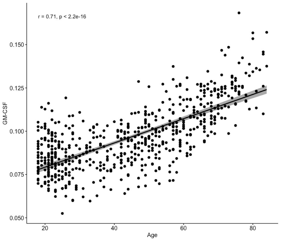
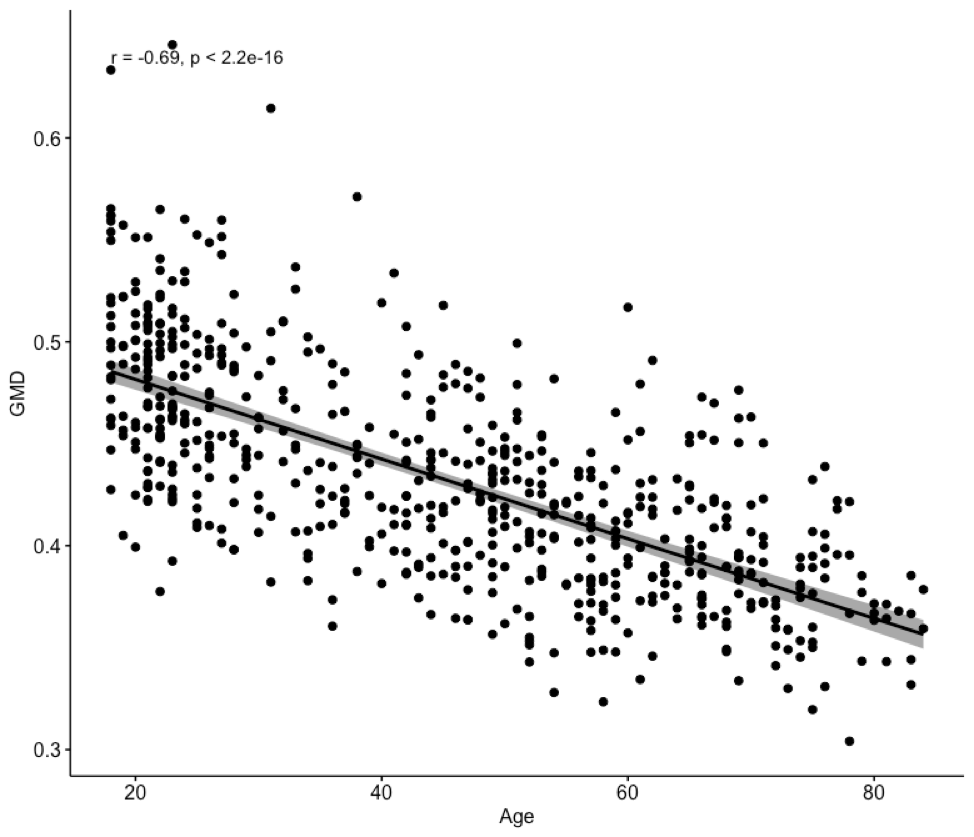
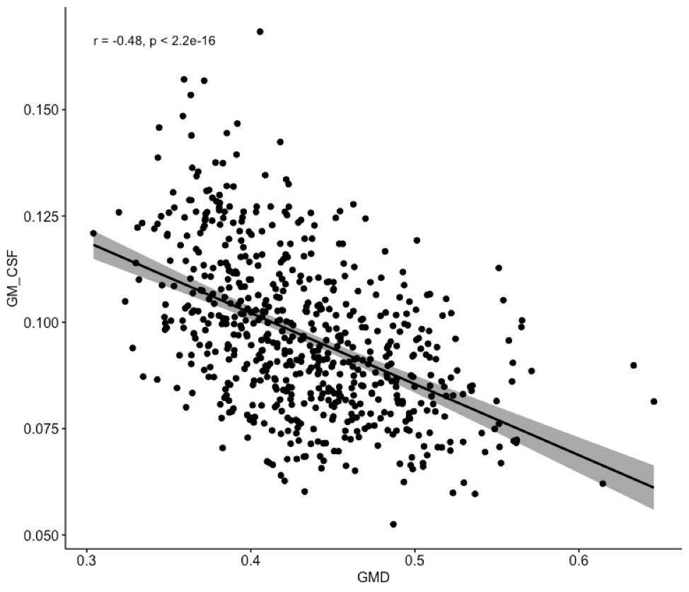
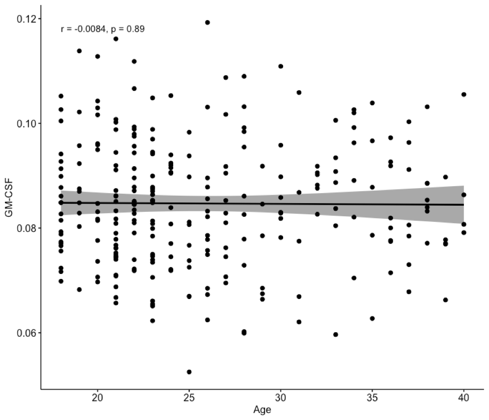
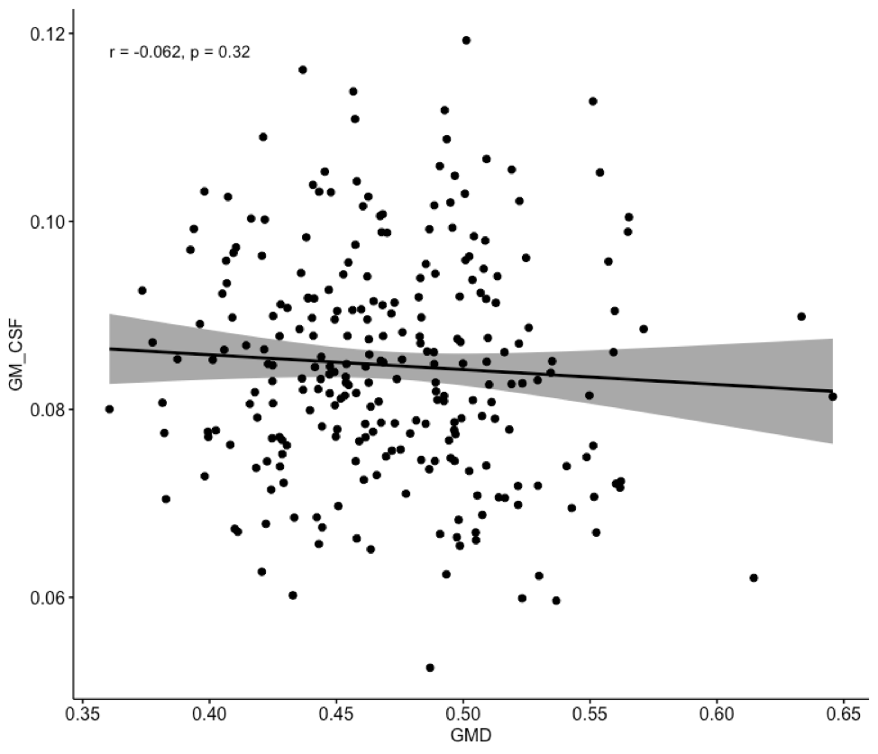
The average CSF signal fraction within grey matter (GM-CSF) showed a strong positive correlation with age (pearson’s r = .71, p<.001) while VBM grey matter density (GMD) showed a strong negative correlation with age (r = -.69, p<.001). Additionally, GM-CSF and GMD were correlated with one another (r = .48, p<.001). To compare the sensitivity of GM-CSF and GMD to early age-related changes in brain structure, the analysis was repeated exclusively with individuals from the cohort younger than 40. GM-CSF was not correlated with age in participants under 40 (r = .008, p = .89), while GMD was negatively correlated with age in participants under 40 (r = -.38, p <.001). GM-CSF and GMD were not correlated with one another in the younger cohort (r = -.062, p = .32).Discussion
GM-CSF and GMD are two methods which can be used to evaluate tissue structure. This study evaluated their relationship to age, and relationship to each other over the lifespan. As predicted, the two methods were negatively related to one another, with GMD decreasing with age and GM-CSF increasing with age. However, perhaps contrary to the original hypothesis, they appear to detect different patterns of change in younger subjects. GMD has a largely linear relationship with age, showing a fairly consistent decrease in grey matter beginning around age 20 and continuing into advanced age. The relationship between GM-CSF and age is non-linear, showing no increase from ages 18-40, and showing fairly linear increases after age 40. It remains unclear why these methods are different in this respect, but it is clear that despite their relationship across a lifespan dataset, the two methods provide different insights into the nature of age-related microstructural changes. Future work will explore the differences in these two techniques and how they may relate to tissue structure.Acknowledgements
No acknowledgement found.References
1. Jeurissen, B., Tournier, J. D., Dhollander, T., Connelly, A., & Sijbers, J. (2014). Multi-tissue constrained spherical deconvolution for improved analysis of multi-shell diffusion MRI data. NeuroImage, 103
2. Dhollander, T., Raffelt, D., & Connelly, A. (2016). Unsupervised 3-tissue response function estimation from single-shell or multi-shell diffusion MR data without a co-registered T1 image. In ISMRM Workshop on Breaking the Barriers of Diffusion MRI (Vol. 5)
3. Mito, R., Dhollander, T., Raffelt, D., Xia, Y., Salvado, O., Brodtmann, A., Rowe, C., Villemagne, V., & Connelly, A. (2018). Investigating microstructural heterogeneity of white matter hyperintensities in Alzheimer’s disease using single-shell 3-tissue constrained spherical deconvolution. Proceedings of the 26th International Society of Magnetic Resonance in Medicine, 26, 0135.
4. Dhollander, T., Raffelt, D., & Connelly, A. (2017). Towards interpretation of 3-tissue constrained spherical deconvolution results in pathology. Proceedings of the 25th International Society of Magnetic Resonance in Medicine, 25, 181
5. Nooner et al, (2012). The NKI-Rockland Sample: A model for accelerating the pace of discovery science in psychiatry. Frontiers in neuroscience 6, 152.
6. Tournier, J. D., Calamante, F., & Connelly, A. (2012). MRtrix: diffusion tractography in crossing fiber regions. International Journal of Imaging Systems and Technology, 22(1), 53-66.
7. Jenkinson, M., Beckmann, C. F., Behrens, T. E. J., Woolrich, M. W., & Smith, S. M. (2012). FSL. NeuroImage, 62(2), 782-790.
8. Avants, B. B., Tustison, N., & Song, G. (2009). Advanced normalization tools (ANTS). Insight j, 2, 1-35.
9. Veraart, J., Fieremans, E., & Novikov, D. S. (2016). Diffusion MRI noise mapping using random matrix theory. Magnetic resonance in medicine, 76(5), 1582-1593.
10. Kellner, E., Dhital, B., Kiselev, V. G., & Reisert, M. (2016). Gibbs‐ringing artifact removal based on local subvoxel‐shifts. Magnetic resonance in medicine, 76(5), 1574-1581.
11. Smith, S. M., Jenkinson, M., Woolrich, M. W., Beckmann, C. F., Behrens, T. E., Johansen-Berg, H., ... & Niazy, R. K. (2004). Advances in functional and structural MR image analysis and implementation as FSL. Neuroimage, 23, S208-S219.
12. Andersson, J. L., Graham, M. S., Zsoldos, E., & Sotiropoulos, S. N. (2016). Incorporating outlier detection and replacement into a non-parametric framework for movement and distortion correction of diffusion MR images. NeuroImage, 141, 556-572.
13. Andersson, J. L., & Sotiropoulos, S. N. (2016). An integrated approach to correction for off-resonance effects and subject movement in diffusion MR imaging. Neuroimage, 125, 1063-1078.
14. Ashburner J. A fast diffeomorphic image registration algorithm. Neuroimage 2007; 38: 95-113.
15. Ashburner J, Friston KJ. Voxel-based morphometry--the methods. NeuroImage 2000; 11: 805-21.
16. Ashburner J, Friston K J. Unified segmentation. NeuroImage 2005; 26, 839-51.
17. Lorio S, Fresard S, Adaszewski S, Kherif F, Chowdhury R, Frackowiak RS., et al. New tissue priors for improved automated classification of subcortical brain structures on MRI. Neuroimage 2016; 130: 157-166.
Figures