2628
Development and evaluation of a 0.5mm isotropic resolution structural template of the older adult brain1Biomedical Engineering, Illinois Institute of Technology, Chicago, IL, United States, 2Rush Alzheimer's Disease Center, Rush University, Chicago, IL, United States
Synopsis
Human brain structural MRI templates with low spatial resolution lack important fine details due to partial volume effects. The purpose of this work was twofold: a) to introduce a novel approach for high-resolution template construction based on principles of super-resolution, and b) using this technique, to develop a high-resolution structural template of the older adult brain based on MRI data from 222 non-demented older adults.
INTRODUCTION
Human brain structural MRI templates with low spatial resolution lack important fine details due to partial volume effects. The purpose of this work was twofold: a) to introduce a novel approach for high-resolution template construction based on principles of super-resolution, and b) using this technique, to develop a high-resolution structural template of the older adult brain based on MRI data from 222 non-demented older adults. The new template was quantitatively compared to other high-resolution templates of the older and younger adult brain, as well as to a lower resolution older adult template, in terms of image quality and in terms of the spatial normalization accuracy achieved when they are used as references for alignment of structural images from a large number of older adults.METHODS
T1-weighted brain MRI data (1mm isotropic) obtained from 222 nondemented older adults (65-95 age-range, male:female=1:1) participating in the Memory and Aging Project1 were used in this work. The proposed method for high-resolution template construction comprised of the following steps:
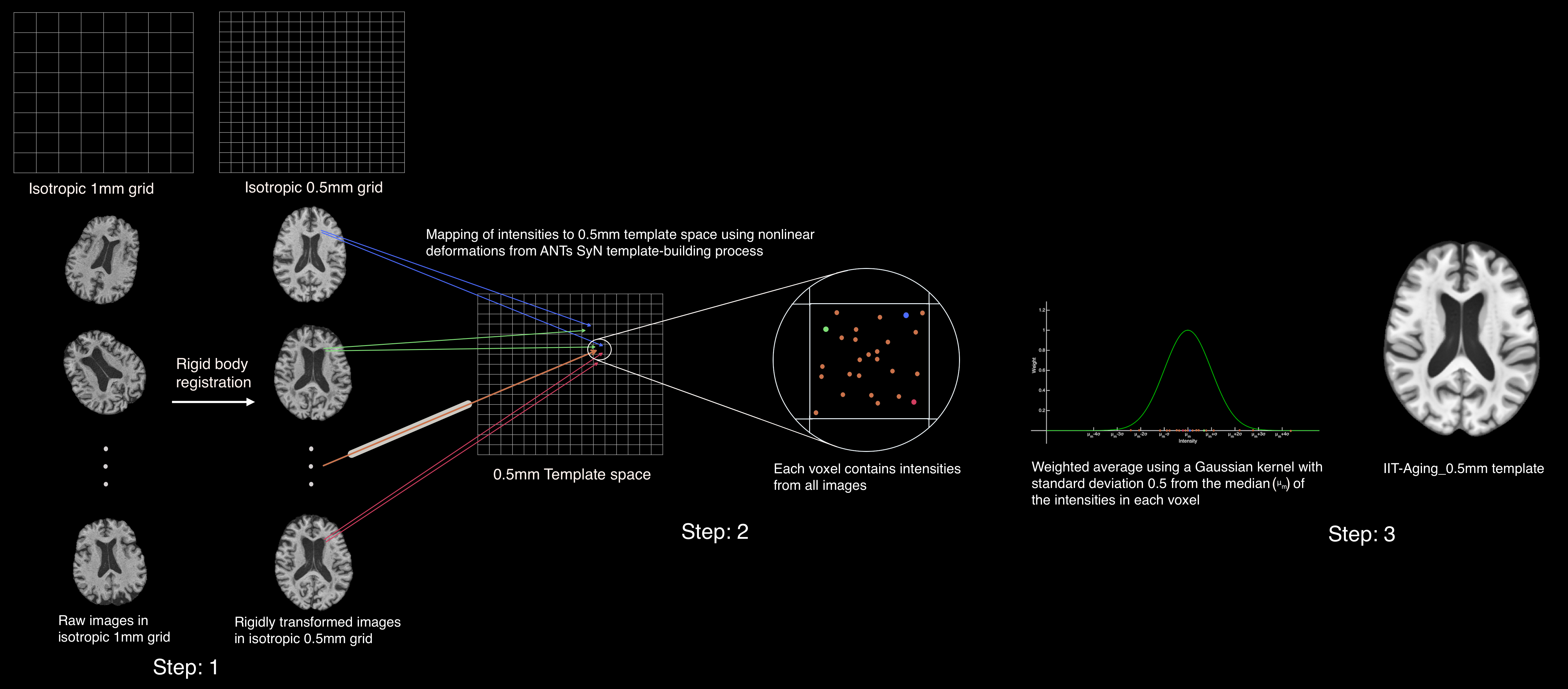
Step 1: Raw images of isotropic 1mm resolution were rigidly and non-linearly aligned in a 0.5mm resolution space using the ANTs2,3 SyN4 based template-building method.
Step 2: The resulting non-linear deformations were utilized to map the image intensities of the rigidly transformed 0.5mm resolution images to exact physical locations (x,y,z) in the 0.5mm template space, eliminating the interpolations that occur in conventional template-building method (Fig.1).
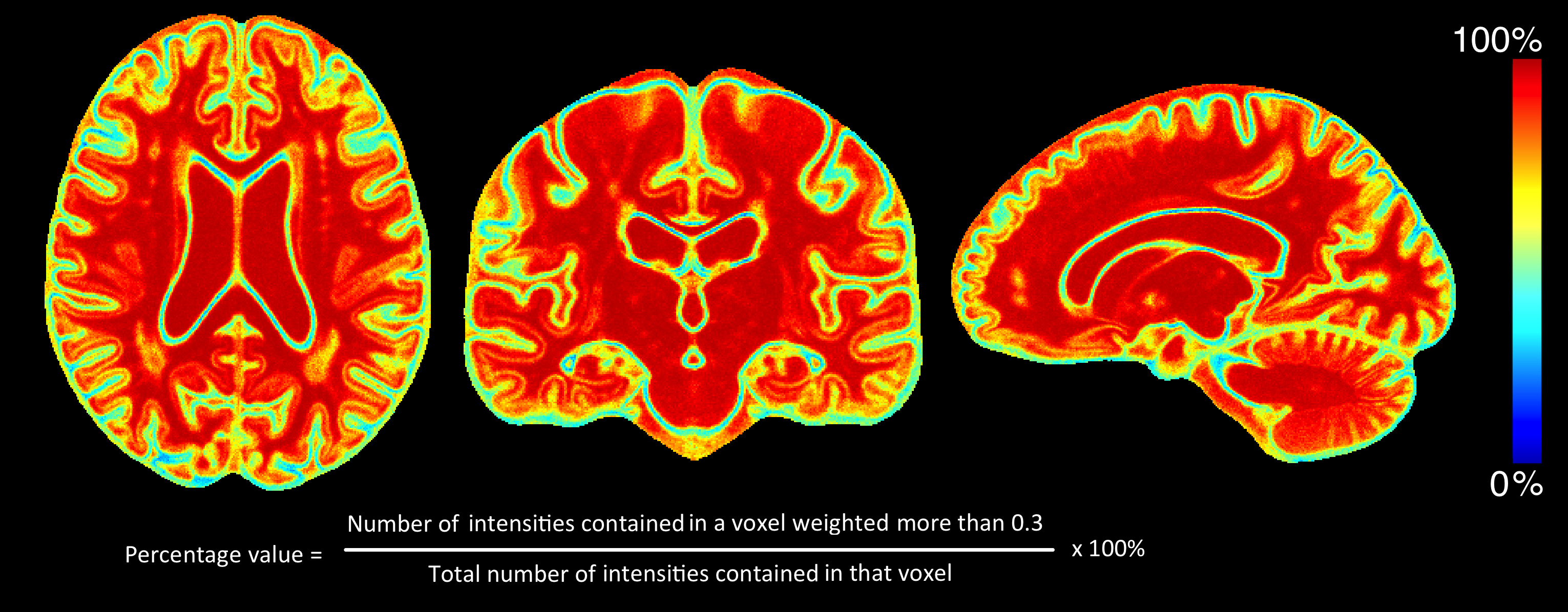
Step 3: The final intensity in each voxel in template space was calculated as the weighted average of the intensities contained in that voxel, using a Gaussian kernel of standard deviation 0.5 from the median of those intensities (Fig.1). This approach is less sensitive to the effects of residual misregistration. Overall, 85% of the voxels in template space had a weight of 0.3 or higher for at least 50% of the intensities included in those voxels (Fig.2) signifying that the majority of the raw signals were utilized in constructing the template.
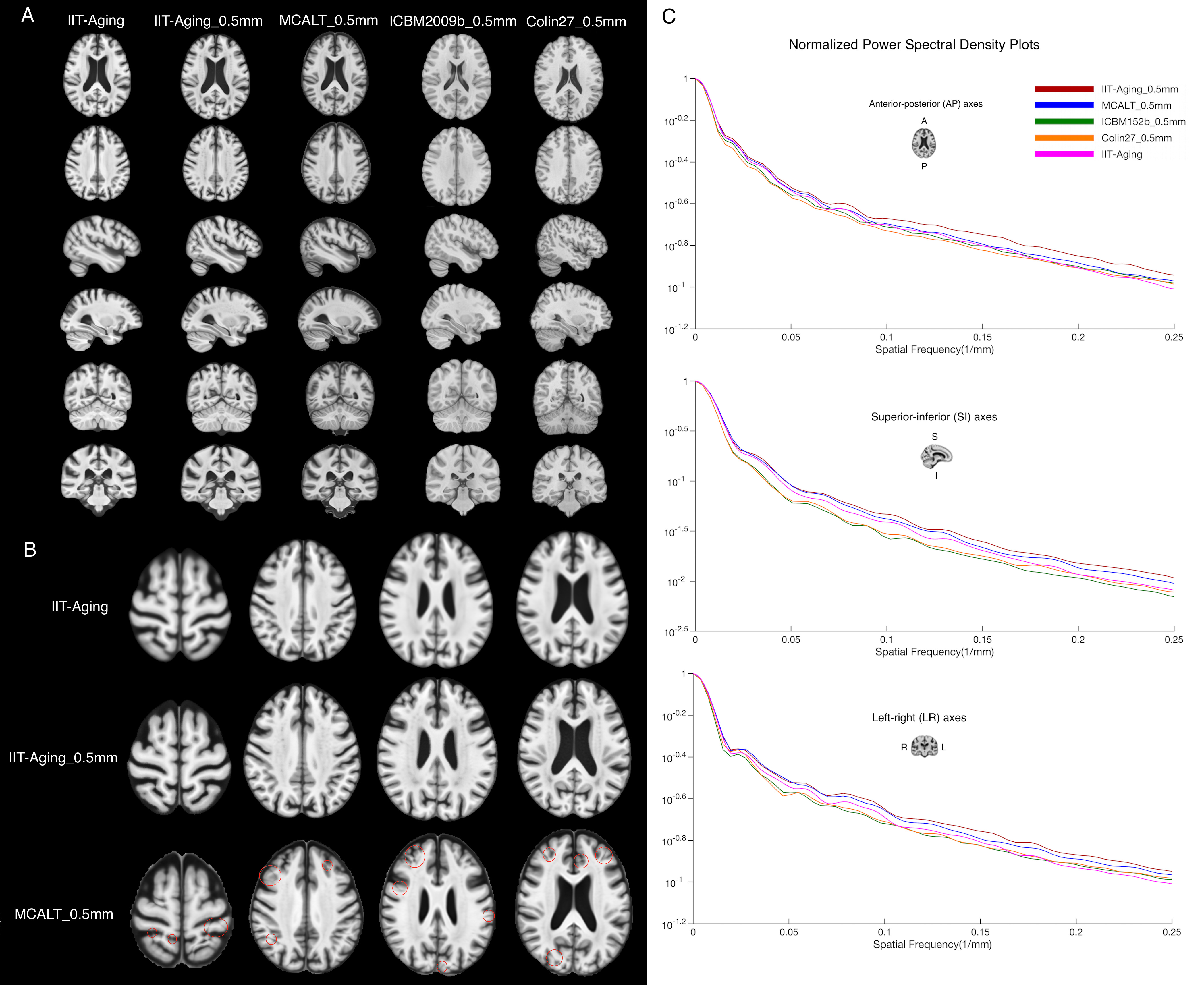
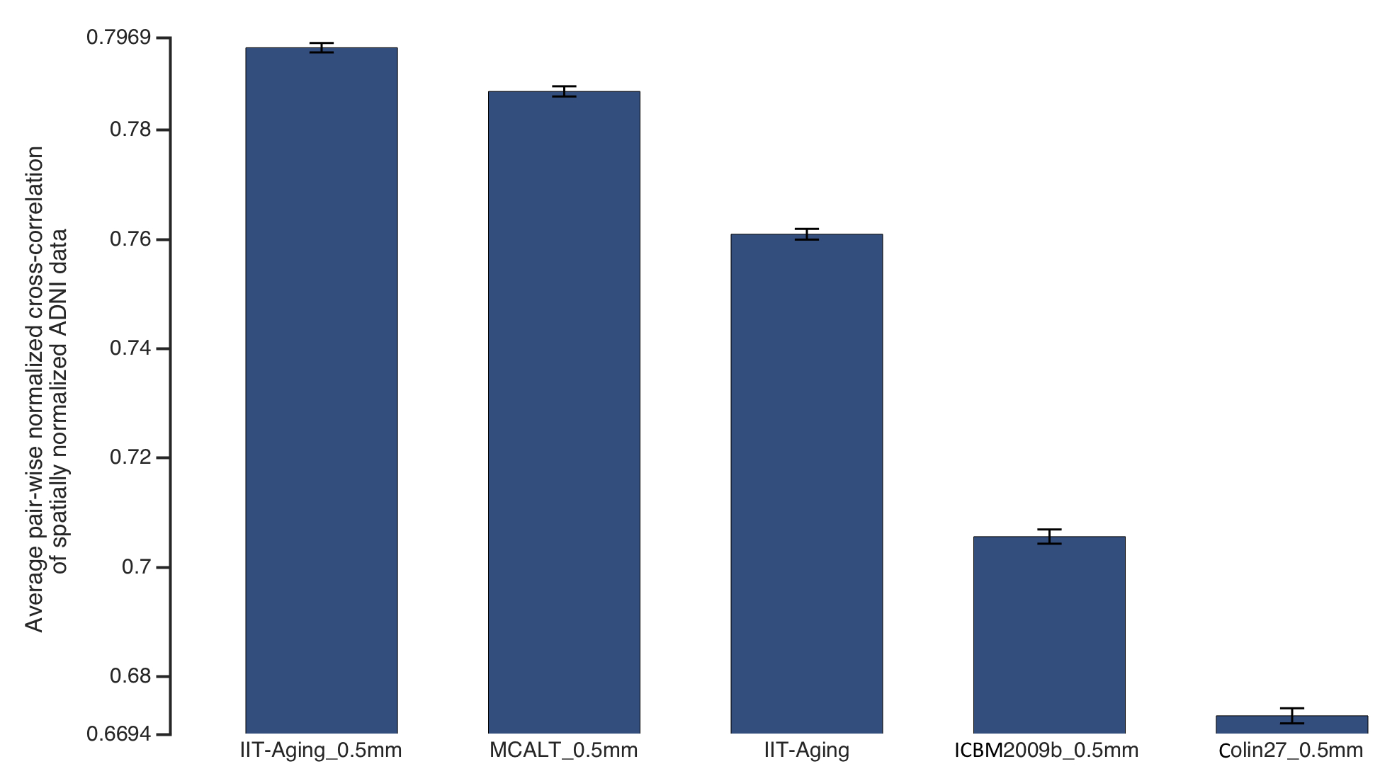
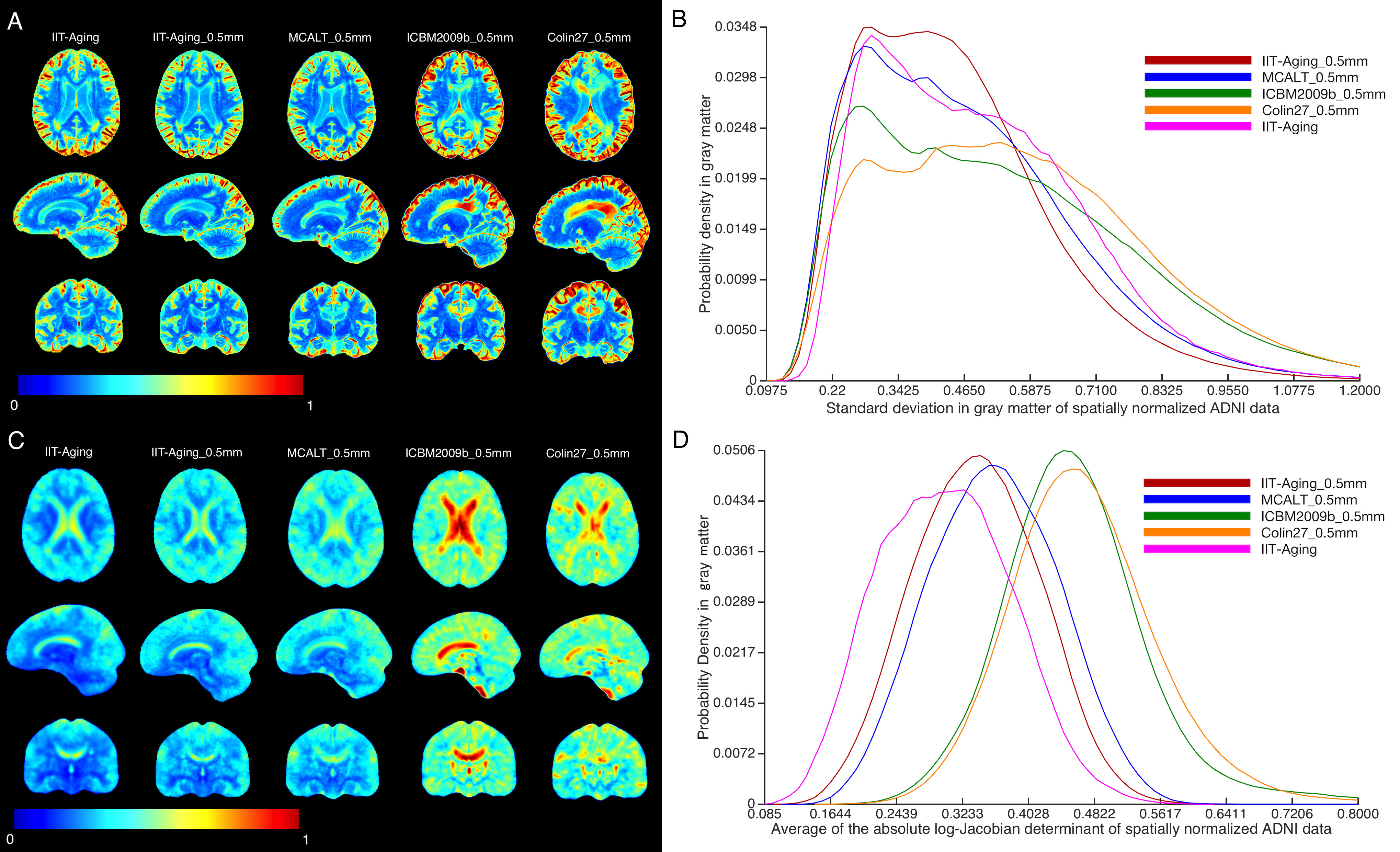
The newly constructed template, referred to as IITAging_0.5mm, was compared to other 0.5mm resolution templates of i) mainly middle-aged and older adults (MCALT_0.5mm5) and ii) young adults (ICBM2009b_0.5mm6,7 and Colin27_0.5mm8), as well as to a recently presented, highly performing, 1mm resolution template constructed from the same raw data using the traditional ANTs SyN template-building method (IIT-Aging9)(Fig.3A). The templates were first compared by visual inspection, and then in terms of image sharpness as demonstrated by the normalized power spectral density, and inter-subject spatial normalization accuracy achieved when used as references for normalization of T1-weighted data (1mm x 1mm x 1.2mm resolution) from 175 non-demented ADNI10 participants. Normalization accuracy was assessed for each template by means of the average pair-wise normalized cross-correlation, standard deviation, and average absolute log-Jacobian determinant in gray matter of the ADNI participants.
RESULTS
The IIT-Aging_0.5mm template has higher image sharpness compared to the other templates, exhibited by larger high spatial frequency content in the normalized power spectra (Fig.3C). Fine structures were resolved in IIT-Aging_0.5mm compared to MCALT_0.5mm and IIT-Aging (1mm resolution) (Fig.3B). Fine features were best resolved in Colin27_0.5mm, however this is a single-subject young adult template, not a population-based older adult template. Visual inspection revealed atypical brain features (artifacts) in the cortex of MCALT_0.5mm (red circles) (Fig.3B), which are not present in the other templates. Finally, the accuracy of inter-subject spatial normalization was higher when using IIT-Aging_0.5mm compared to the other templates (Fig.4, Figs.5A,B). The IIT-Aging and IIT-Aging_0.5mm templates required lower spatial deformation for normalizing ADNI data (Fig.5C, Fig.5D).DISCUSSION
The new IIT-Aging_0.5mm template is a population-based template of the older adult brain that exhibits high image quality, high sharpness, is free of artifacts, and resolves fine brain structures. The IIT-Aging_0.5mm template provided higher spatial normalization accuracy of ADNI data compared to the 0.5mm resolution, middle-aged and older adult template MCALT_0.5mm. The improvement in spatial normalization achieved with IIT-Aging_0.5mm was even more substantial when compared to the 0.5mm resolution, young adult templates (ICBM2009b_0.5mm, Colin27_0.5mm). This demonstrates the importance of using population-based, older adult templates in studies of aging. Finally, the improvement in spatial normalization achieved with IIT-Aging_0.5mm compared to IIT-Aging demonstrates that the higher quality and higher resolution of IIT-Aging_0.5mm is advantageous even for spatial normalization of lower resolution ADNI data.CONCLUSION
The findings of this work have important implications in regard to template selection in studies of older adults. The IIT-Aging_0.5mm template is a high-quality, high-resolution structural template of the older adult brain that provides higher spatial normalization accuracy than other templates of the older as well as younger adult brain, even for normalization of lower resolution older adult data.Acknowledgements
National Institute on Aging R01AG052200References
1. A Bennett D, A Schneider J, S Buchman A, et al. Overview and findings from the rush Memory and Aging Project. Current Alzheimer Research. 2012;9(6):646-63.
2. Avants BB, Yushkevich P, Pluta J, et al. The optimal template effect in hippocampus studies of diseased populations. Neuroimage. 2010;49(3):2457-66.
3. Avants BB, Tustison NJ, Song G, et al. A reproducible evaluation of ANTs similarity metric performance in brain image registration. Neuroimage. 2011;54(3):2033-44.
4. Avants BB, Epstein CL, Grossman M, et al. Symmetric diffeomorphic image registration with cross-correlation: evaluating automated labeling of elderly and neurodegenerative brain. Medical image analysis. 2008;12(1):26-41.
5. Schwarz CG, Gunter JL, Ward CP, et al. THE MAYO CLINIC ADULT LIFE SPAN TEMPLATE: BETTER QUANTIFICATION ACROSS THE LIFE SPAN. Alzheimer's & Dementia: The Journal of the Alzheimer's Association. 2017;13(7):P93-4.
6. Fonov V, Evans AC, Botteron K, et al. Brain Development Cooperative Group. Unbiased average age-appropriate atlases for pediatric studies. Neuroimage. 2011;54(1):313-27.
7. Fonov VS, Evans AC, McKinstry RC, et al. Unbiased nonlinear average age-appropriate brain templates from birth to adulthood. NeuroImage. 2009(47):S102.
8. Holmes CJ, Hoge R, Collins L, et al. Enhancement of MR images using registration for signal averaging. Journal of computer assisted tomography. 1998;22(2):324-33.
9. Ridwan AR, Zhang S, Qi X, et al. EVALUATION OF STANDARDIZED T1-WEIGHTED BRAIN TEMPLATES FOR USE IN STUDIES ON OLDER ADULTS. Alzheimer's & Dementia: The Journal of the Alzheimer's Association. 2018;14(7):P852-3.
10. Weiner MW, Veitch DP, Aisen PS, et al. The Alzheimer's Disease Neuroimaging Initiative: a review of papers published since its inception. Alzheimers Dement. 2013;9(5):e111-94.
Figures