2624
Development of a Standardized Normative Pediatric Spinal Cord structural template: Demonstration of an automatic estimation of Spinal Cord Cross Sectional Area measurements (SCCSA).Shiva Shahrampour1, Benjamin De Leener2, Devon Middelton3, Kavya Jonnavithula4, Mahdi Alizadeh5, Hiba F Pediyakkal6, Laura Krisa7, Adam Flanders8, Scott Faro9, Julien Cohen-Adad2, and Feroze Mohamed7
1Bioengineering, Temple University, Philadelphia, PA, United States, 2Electrical Engineering, NeuroPoly Lab, Institute of Biomedical Engineering, Montreal, QC, Canada, 3Radiology, Thomas Jeffesron University, Philadelphia, PA, United States, 4School of Biological Sciences, University of California, Irvine, Irvine, CA, United States, 5Neurosurgery, Thomas Jefferson University, Philadelphia, PA, United States, 6Department of Chemistry, Boston University, Boston, MA, United States, 7Thomas Jefferson University, Philadelphia, PA, United States, 8Radiology, Thomas Jefferson University, Philadelphia, PA, United States, 9School of Medicine, Johns Hopkins, Baltimore, MD, United States
Synopsis
Template-based analysis of MRI data of the spinal cord lay the foundation for standardization and reproducibility , improves patient diagnosis and helps the discovery of new biomarkers of spinal-related diseases.
Purpose:
The purpose of this work is to create a structural MRI based template of the normal pediatric spinal cord by combining T2-weighted MR scans of several typically developing subjects. This will allow clinicians and researchers to objectively evaluate and quantify various structures within the spinal cord. In this current work we have also shown how this template can be utilized to quantify the spinal cord cross section area (SCCSA).Materials and method:
A T2-weighted 3D SPACE sequence from 30 typically developing (TD) pediatric subjects ranging in age from 6-16 yrs old was acquired to cover C1-T12 vertebrae in two slabs. The slabs were stitched into a single volume to exhibit the entire cord using the vendor provided software on the scanner. The scans were performed using a 3.0T Siemens Verio MR scanner and the imaging parameters were: voxel size = 1×1×1 mm3, TR=1500 ms, TE=122 ms and Slice thickness=1 mm. Several pre-processing steps were performed on MRI images on all the 30 subjects before the actual template generation as described below: (I) spinal canal centerline extraction; was performed on all images to accurately segment the spinal canal starting from the edge of the delineated brainstem. (II) The position of intervertebral discs was then semi-automatically identified using a template-matching detection algorithm [1] and (III) a slice-based intensity normalization procedure was applied to all images to normalize image intensity of the inside of spinal cord to the average intensity of the entire dataset. After successful pre-processing, the final pediatric template was created using the following pipeline. (I) Initially the spinal cord centerline, and the intervertebral discs positions were semi-automatically extracted on all images using PropSeg (sct_propseg) [2] and vertebral labeling (sct_label_vertebrae) tools [1] from Spinal Cord Toolbox (SCT) [3]. (II) Next the spinal cord was straightened, and vertebral levels were aligned using a Non-Uniform Rational Bezier Spline (NURBS) based nonlinear transformation [4]. (III) Finally, an unbiased left-right symmetric template was constructed using a hierarchical group-wise image-registration method [5]. The image registration method used in this procedure is based on the nonlinear registration engine of Automatic Nonlinear Image Matching and Anatomical Labeling (ANIMAL) [6]. The template generation algorithm computes the average of all subjects iteratively and registers the images to this average nonlinearly. As a demonstration of the utilization of this template, spinal cord cross sectional area (SCCSA) was computed at all the disc levels in all the subjects of this study and is compared to SCCSA measured in the generated template, Fig (2). The comparison is also used as sanity-check to confirm that the nonlinear deformations applied to images during template generation preserves the topology of spinal cord, as previously verified on adult spinal cords [4]. The Intra Correlation Coefficient (ICC) is also computed as 0.94 for selected disc levels as shown in Table (1). This measure, quantifies the intra SCCSA measurements reliability between the two groups as well as the consistency of the measurements in two groups relative to one another.Result:
Fig (1) shows
sagittal and coronal view of the template along with the labeled vertebral
bodies and segmented cord. Axial views illustrate probabilistic map of white
and gray matter and Cerebrospinal Fluid (CSF) as well as cord segmentation. Fig
(2) top window shows the SCCSA of the produced pediatric template (average
across levels: 46.5 mm2 ±12.5) and the bottom window shows the
average SCCSA across all 30 subjects (average across levels: 47 mm2
±9.6). The similarity in the shapes of the plots suggests the intactness of the
overall structure of the spinal cord after straightening and deformation
process during template registration. Table (1) shows the SCCSA measurements
along with Coefficient of Varian (CV) for selected disc levels. The ICC of
0.94, indicates a strong agreement between the measurements in both groups.Conclusion:
To
the best of our knowledge this work is the first to create a standardized
template of spinal cord in pediatric subjects. As described, a method is presented for pediatric template generation
which is unbiased to subject selection and preserves the topology of the spinal
cord. Utility of this template in automatically estimating the SCCSA is also
demonstrated. Future work with a larger cohort with varied age ranges and gender
is warranted.Acknowledgements
No acknowledgement found.References
[1]: Ullmann, E., Pelletier Paquette, J., Thong, W., and Cohen-Adad, J., 2014, "Automatic Labeling of Vertebral Levels Using a Robust Template-Based Approach", International Journal of Biomedical Imaging, 2014, pp. 1-9. [2]: De Leener, B., Kadoury, S., and Cohen-Adad, J., 2014, "Robust, accurate and fast automatic segmentation of the spinal cord", NeuroImage, 98, pp. 528-536. [3]: De Leener, B., Lévy, S., Dupont, S., Fonov, V., Stikov, N., Louis Collins, D., Callot, V., and Cohen-Adad, J., 2017, "SCT: Spinal Cord Toolbox, an open-source software for processing spinal cord MRI data", NeuroImage, 145, pp. 24-43. [4]De Leener, B., Mangeat, G., Dupont, S., Martin, A., Callot, V., Stikov, N., Fehlings, M., and Cohen-Adad, J., 2017, "Topologically preserving straightening of spinal cord MRI", Journal of Magnetic Resonance Imaging, 46(4), pp. 1209-1219. [5]: Fonov, V., Le Troter, A., Taso, M., De Leener, B., Lévêque, G., Benhamou, M., Sdika, M., Benali, H., Pradat, P., Collins, D., Callot, V., and Cohen-Adad, J., 2014, "Framework for integrated MRI average of the spinal cord white and gray matter: The MNI–Poly–AMU template", NeuroImage, 102, pp. 817-827. [6]: Collins, D., Holmes, C., Peters, T., and Evans, A., 1995, "Automatic 3-D model-based neuroanatomical segmentation", Human Brain Mapping, 3(3), pp. 190-208.Figures
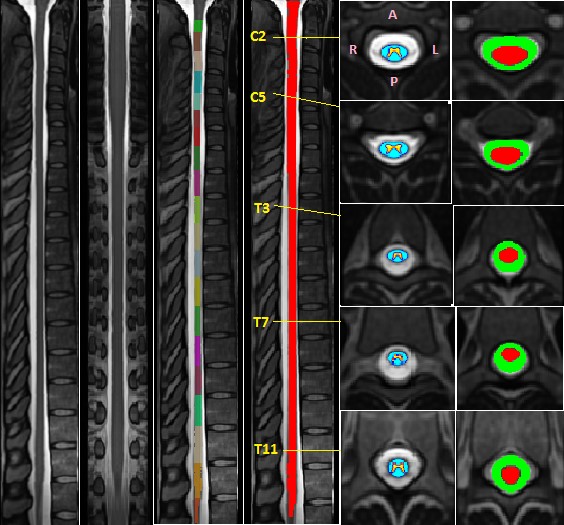
Figure (1): The left four images show the sagittal and
coronal view of the template along with the vertebral labels and segmented cord
in red. The right two images show probabilistic map of white and gray matter as
well as Cerebrospinal Fluid (CSF) and cord segmentation.
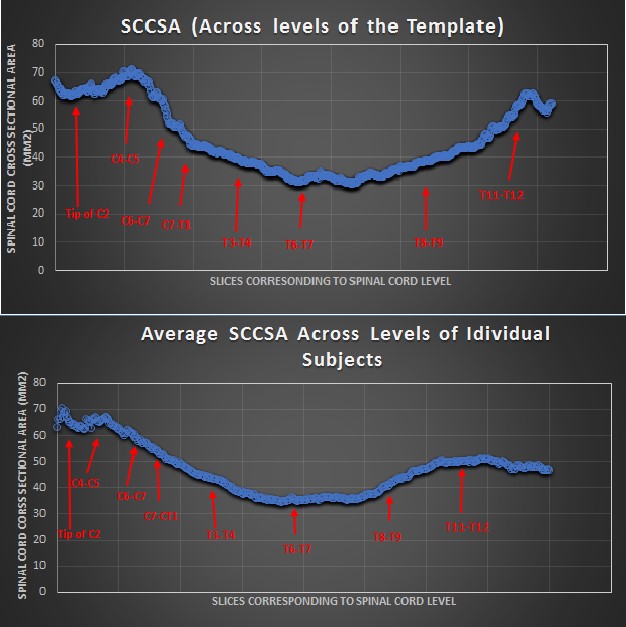
Figure
2: Top window shows the SCCSA of the pediatric template (average across levels:
46.5 mm2 ±12.5). The bottom window shows average SCCSA across all 30 subjects
(average across levels: 47 mm2 ±9.6). This suggests that SCCSA is preserved
between native subjects’ space and the template.
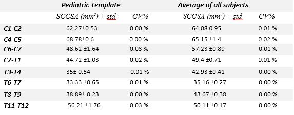
Table 1: Comparison of SCCSA (mm2) along
with Coefficient of Variation (CV) in selected intervertebral discs.