2619
Altered brain structure associated with cognitive changes of end-stage renal disease patients without dialysis and with maintenance hemodialysis1Medical Imaging department, The First Affiliated Hospital of Xi'an Jiaotong University, Xi'an, China
Synopsis
The theory of kidney-brain axis has provided more information for the interpretation of brain damage in ESRD patients. However, the factor of dialysis was ignored in this theoretical system. We analyzed the cortical structural changes and cognitive changes from different dimensions and also analyzed their relationship in ESRD patients with and without hemodialysis. We found that both the patients with dialysis and the patients without dialysis showed decreased cortical thickness when compared with healthy people, while the patients without dialysis presented with a more extensive decreased cortical thickness when compared with patients with maintenance hemodialysis. The brain structural changes were correlated with the cognitive changes. Our results suggested that the hemodialysis might be a protective factor for the brain, but the protective effect of hemodialysis was limited.
Introduction
End-stage renal disease (ESRD), as the last stage of chronic kidney disease, is characterized by an estimated glomerular filtration rate (eGFR) less than 15 ml/min/1.73 m2 and the need for renal replacement therapy via dialysis and transplantation1. The brain is one of main injured organs in the development of ESRD, and patients often present with stroke, sensorimotor abnormality and cognitive dysfunction. The cognitive impairment was reported affected up to 70% ESRD patients and could complicate ESRD treatment and increase the mortality 2-4. More and more researchers have suggested that the brain injury in ESRD patients might be attributed to the cerebrovascular damage and neurodegenerative changes induced by many traditional and non-traditional risk factors and uremic toxins 5. However, several studies also indicated that the hemodialysis procedure played an important role in the neuropathological changes in ESRD patients 6,7. Thus, the brain structure or function evaluation in ESRD patients before dialysis initiation is very essential for the uncovering of the mechanisms underlying brain damage. Here, we focused on exploring the different brain injury patterns and the relationships between brain changes and cognitive impairment in ESRD patients with and without hemodialysis.Methods
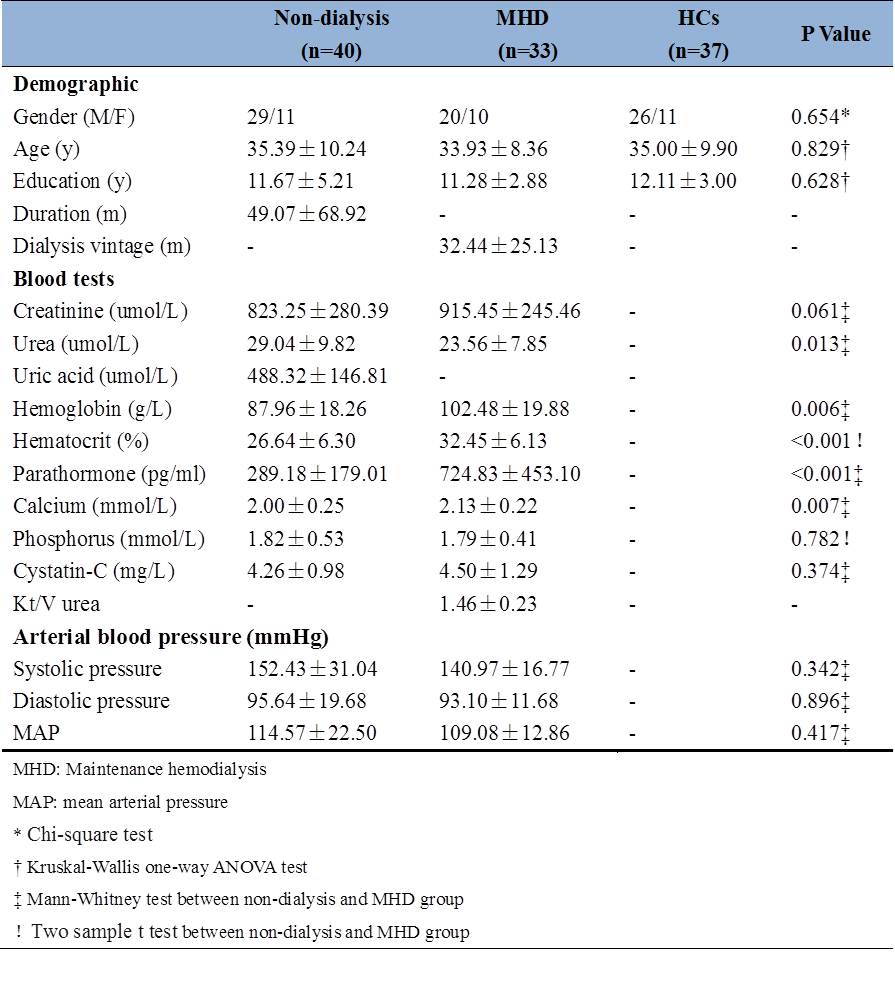
40 ESRD patients without dialysis (mean age 35.39±10.24 years) and 33 maintenance hemodialysis patients and (mean age 33.93±8.36 years) and 37 healthy volunteers (mean age 35.00±9.90 years) were included in our study. Three-dimensional T1-weighted imaging was acquired using a GE 3.0T HDxt scanner. Cortical thickness was calculated used FreeSurfer software (http://surfer.nmr.mgh.harvard.edu/). All the subjects were evaluated global cognitive function by Montreal Cognitive Assessment (MoCA), episodic memory function by Auditory Verbal Learning Test (AVLT), executive and attention function by Trail Making Test A, and working memory by a computed based n-back working memory battery. We collected the laboratory blood tests in all ESRD patients (table1).Results
The ESRD patients without dialysis had a lower level of hemoglobin, hematocrit, parathromone and calcium when compared with maintenance hemodialysis patients (the hemodialysis patients were treated with erythropoietin) (table1). Both ESRD patients with and without hemodialysis showed lower scores in MoCA and longer time in Trail Making Test A when compared with healthy controls (P<0.05). For memory test, ESRD patients without hemodialysis presented with lower scores than both hemodialysis patients and controls (P<0.05). For the working memory test, the patients without dialysis showed increased reaction time (P<0.05) (table2).Compared with healthy controls, the ESRD patients without dialysis showed extensive decreased cortical thickness, including bilateral postcentral gyrus, precentral gyrus, superior frontal gyrus, superior temporal gyrus, inferior parietal lobule and precuneus (P<0.01, FDR corrected). The maintenance hemodialysis patients also presented with decreased cortical thickness compared with healthy controls, but effected regions were relatively smaller than ESRD patients without dialysis, mainly including bilateral postcentral gyrus, right inferior frontal gyrus, and bilateral inferior parietal lobule (P<0.01, FDR corrected). When compared the cortical thickness of two ESRD patients group, the patients without dialysis presented with widespread decreased thickness, mainly including the bilateral postcentral gyrus, prefrontal cortex, and inferior parietal lobule (P<0.01, FDR corrected) (figure1). The changes of thickness of right inferior parietal lobule, right postcentral gyrus, right precuneus were positively correlated with hemoglobin level (P<0.01, FDR corrected). The correlation between cortical structure changes and cognition showed that the memory and executive function changes was correlated with the thickness changes (figure2).Discussion
Previous studies showed the single dialysis could improve the cognitive function, but also studies suggested that the dialysis was a factor that could accelerate the decline of cognitive function in ESRD patients 6,7. Our results showed that maintenance patients showed a better cognitive performance and brain structure, which indicated that the hemodialysis played a possible protective role of the brain. The hemodialysis procedure could remove excessive organic osmolytes and brain water which might relieve the microstructural edema. Another possible explanation was the treatment with erythropoietin to correct the renal anemia in hemodialysis patients. Low hemoglobin tended to reduce both blood viscosity and oxygen supply, which would result in the hypoxia of brain tissue8. This study might help us better understand the pathophysiology of neural damage patterns in ESRD patients.Conclusion
Compared with ESRD patients without dialysis, maintenance hemodialysis patients presented with improved brain structure and function accompanied with improved cognition, which indicated that the hemodialysis might be a protective factor for the brain. However, when compared healthy people, the decreased brain structure and function were found no matter in ESRD patients without dialysis or maintenance hemodialysis patients, which indicated that the protective effect of hemodialysis was limited.Acknowledgements
This research was supported by the National Natural Science Foundation of China (Grant No. 81371530 and 81571640) and Natural Science Foundation of Shaanxi Province of China (Grant No. 2017ZDJC-13).References
- Foley RN, Collins AJ. End-stage renal disease in the United States: an update from the United States Renal Data System. J Am Soc Nephrol 2007;18(10):2644-8.
- Dong J, Pi HC, Xiong ZY, Liao JL, Hao L, Liu GL, et al. Depression and Cognitive Impairment in Peritoneal Dialysis: A Multicenter Cross-sectional Study. American journal of kidney diseases: the official journal of the National Kidney Foundation. 2016;67(1):111-8.
- McQuillan R, Jassal SV. Neuropsychiatric complications of chronic kidney disease. Nat Rev Nephrol 2010;6(8):471–479.
- Harhay MN, Xie D, Zhang X, Hsu CY, Vittinghoff E, Go AS, et al. Cognitive Impairment in Non-Dialysis-Dependent CKD and the Transition to Dialysis: Findings From the Chronic Renal Insufficiency Cohort (CRIC) Study. American journal of kidney diseases: the official journal of the National Kidney Foundation. 2018;72(4):499-508.
- Bugnicourt JM, Godefroy O, Chillon JM, Choukroun G, Massy ZA. Cognitive disorders and dementia in CKD: the neglected kidney-brain axis. Journal of the American Society of Nephrology: JASN. 2013;24(3):353-63.
- Lux S, Mirzazade S, Kuzmanovic B, Plewan T, Eickhoff SB, Shah NJ, et al. Differential activation of memory-relevant brain regions during a dialysis cycle. Kidney international. 2010;78(8):794-802.
- Li P, Ding D, Ma XY, Zhang HW, Liu JX, Zhang M. Altered intrinsic brain activity and memory performance improvement in patients with end-stage renal disease during a single dialysis session. Brain imaging and behavior. 2018.
- Sun YY, Lee J, Huang H, Wagner MB, Joiner CH, Archer DR, et al. Sickle Mice Are Sensitive to Hypoxia/Ischemia-Induced Stroke but Respond to Tissue-Type Plasminogen Activator Treatment. Stroke. 2017;48(12):3347-55.
Figures