2617
Altered cortical thickness relevance in the early blind, late blind during the critical developmental time1Neurology, All India Institute of Medical Sciences, New Delhi, India, 2NMR and MRI Facility, All India Institute of Medical Sciences, New Delhi, India, 3Rajendra Prasad Centre of Opthalmology, All India Institute of Medical Sciences, New Delhi, India
Synopsis
Visual impairment induces
introduction
Blindness is allied with established morphometry and function changes of the occipital cortex [1]. Visual impairment induces the structural and functional alteration in the primary visual cortex (V1). Studies reported the early blind subjects had a thicker occipital cortex compared to late blind and sighted controls [1,2]. Hence onset of blindness and age of blindness may play a significant role changes of cortical thickness occipital cortex.Objective
To observe the structural alteration by mapping cortical thickness in the visual cortex in early blind, late blind and sighted control groups of age range 6 to12 years and 13 to 19 yearsMethodology
Ten early blind (EB) and ten late blind (LB) subjects and ten sighted controls (SC) (all right-handed) in two age groups 6-12 years and 13-19 years were recruited from the clinics of our institute (Table 1). Standard diagnostic and exclusion criteria were followed. T-1 weighted data were acquired using 3T MR scanner (Achieva3.0T TX, Philips, Netherlands), with the following imaging parameters: slices per slab: 160, distance factor 50%, orientation: sagittal; slice thickness: 1mm; T1:1100ms; TR: 1900ms; TE: 3.37ms; averages: 1; FOV: 256mm, FOV phase: 93.8%; Base resolution: 256; Phase resolution 100; Phase encoding direction: A>>P; Bandwidth: 130 Hz; echo spacing: 8.6ms.. Pre- and post-processing were carried out using SPM12 with the help of CAT12 toolbox (The Wellcome Department of Cognitive Neurology, University College, and London, UK).). The clusters were converted from MNI template to the Talairach and Tornoux coordinates, for estimation of anatomical areas [3]. One-way ANOVA (p<0.001, cluster threshold 10) was used for group analyses.Result
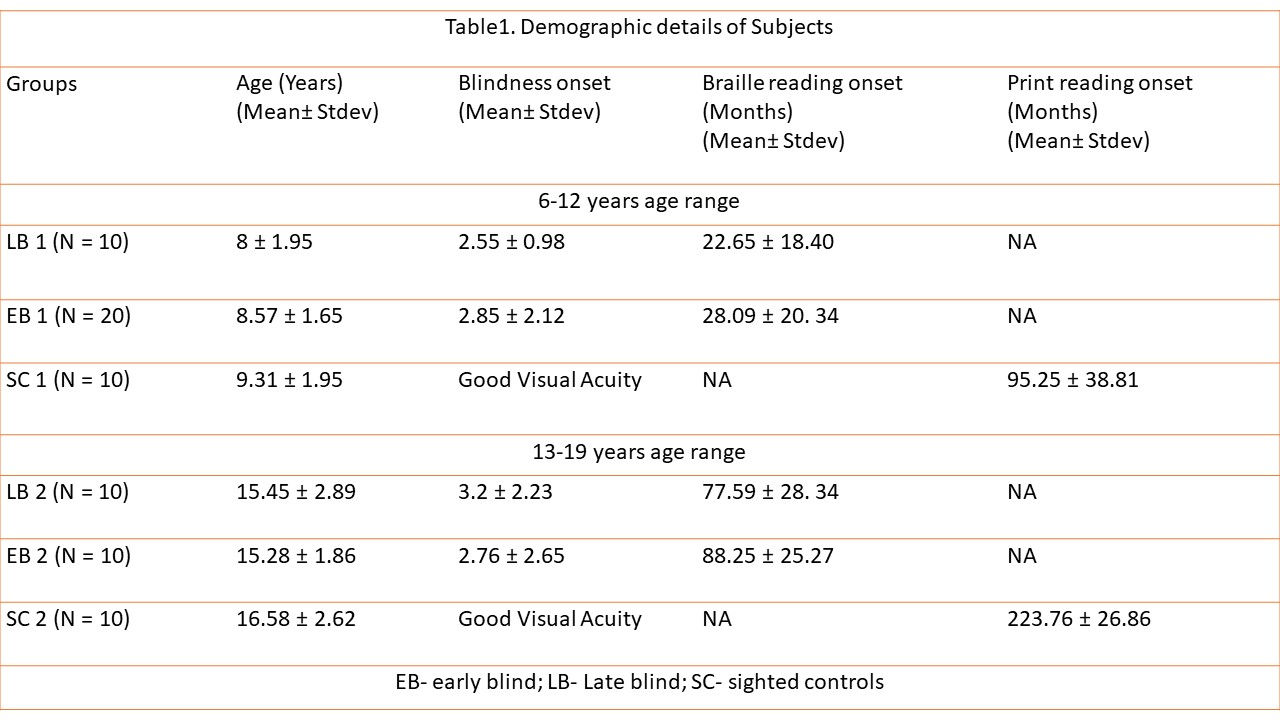
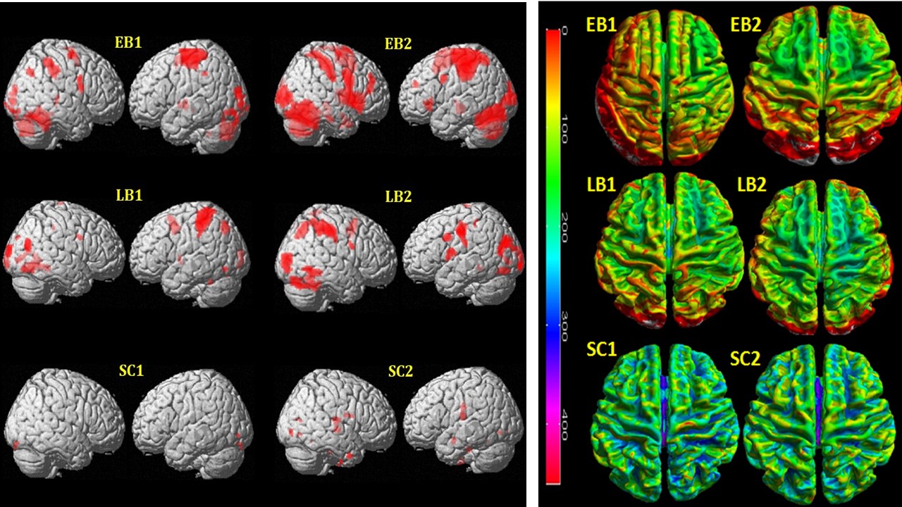
Cortical thickness (CT) measures in blind (early and late) and sighted subjects were obtained (Figure 1). The most remarkable finding was that the early blind had a thicker cortex at the bilateral primary visual cortex and left visual association cortex than the sighted controls (p < 0.05 FWE, extent threshold k=10). Group contrasts confirmed previous results in showing thicker occipital cortex in the EB1< EB2 and LB2 both groups (13-19 years age range). However, there was no difference in the LB1 and Sighted control groups (SC1 and SC2).Discussion
Increased cortical thickness of occipital areas in early blind of age range 6-12 and 13-19 years along with late blind 13-19 years subjects may be indicating improvement of other sensory modalities (tactile and haptic) induce behavioural enhancements. These results are reflecting cross-modal plasticity due to adaptive compensatory mechanism in brain of visually deprived subjects. On comparing the EB with the LB, results showing effects the age of onset of blindness and the total duration of blindness may have playing role in cortical thickness [4]Conclusion
Cortical thickness revealing structural changes are dependent on onset of blindness and age of blindness.Acknowledgements
Department of NMR and MRI facility and DST for funding the study.References
1. Atilgan H et al Brain Lang. 2017 Jul;170:71-81.
2. Jiang J et al The Journal of Neuroscience, 2009 • 29(7):2205–2211 • 2205
3. Talairach J, and Tornoux P 1988 Stuttgart: thieme
4. VosP et al Cerebral Cortex 2012;22:2455– 2465
Figures