2612
Plastic Changes of the Language-related Brain Regions for Children with Non-syndrome Cleft of Lip with or without Palate (NSCL/P)1Beijing Children’s Hospital, Capital Medical University, beijing, China, 2MR Research China, GE Healthcare, beijing, China, 3Beijing Stomatological Hospital, Capital Medical University, beijing, China, 4Beijing Normal University, beijing, China
Synopsis
Using multimode MRI technique, this study attempt to find structural and functional alterations of brain regions for children with non-syndrome cleft of lip with or without palate (NSCL/P). Compared with control group, both structural and functional changes were detected in distributed cortical regions for NSCL/P group, which mainly located on the dorsal stream of language pathways. Besides, significant correlations were found between ALFF values and Chinese language clear degree scales for NSCL/P children.
Introduction
Neuroplasticity is well known as that brain owned an amazing ability to make structural and functional plastic changes in reaction to behavioral experiences, environmental stimulus and cognitive demands.1-3 Multimode MR imaging provided feasible methods to detect both structural and functional plastic alterations of the brain in vivo. With velopharyngeal insufficiency, the NSCL/P children practice in aberrant articulation behaviors trying to close the velopharyngeal cavity and prevent nasal leakage. It may result in abnormal alterations of nasality, formant of vowel, the values of jitter, harmonics-to-noise ratio and cepstral prominence peaks.4 We hypothesis the aberrant phonology cognition and articulation behavior may induce the plastic changes in language-related cortical regions. The purpose of this study is to explore whether there are structural and functional alterations in brain regions for NSCL/P children using multimode MR imaging.Methods
A total of 25 children (4.8-15.4yrs) with NSCL/P and 25 gender- and age-matched healthy controls underwent 3D T1 weighted images and rs-fMRI examinations on a 3.0 T MR scanner. Preprocessing steps were conducted by using the CAT12 (http://dbm.neuro.uni-jena.de/cat/) and DPARSFA (http://www.restfmri.net/forum/) software. The parameters of the surface-based morphometry including cortical thickness (CT), gyrification index (GI), sulcus depth and fractal dimension were computed based on 3D T1 images. In addition, the parameters of local spontaneous brain activities were calculated based on rs-fMRI images, such as regional homogeneity (ReHo), amplitude of low frequency fluctuations (ALFF) and fractional ALFF (fALFF). Between-group differences of computed parameters were assessed with a series of two-sample t-tests. The threshold was set as Gaussian Random Field (GRF) theory corrected p < 0.05. Besides, Chinese language clear degree scale (CLCDS) were performed in NSCL/P group and the correlations between values of all parameters and CLCDS were calculated.Result
Significant between-group differences were detected in distribution cortical regions for both structural (CT, GI) and functional (ALFF) parameters (see Fig.1). Cortical regions with significant structural alterations all located in the left hemisphere. The regions with increased CT values included the operculum cortex, the precentral and postcentral gyrus, the inferior/superior frontal gyrus, the middle/lateral occipital cortex, the frontal orbital cortex and frontal pole, the superior temporal gyrus, the planum polare and the fusiform (Fig.1 a). And the regions with increased GI values located on the middle of the precentral gyrus, the inferior of the postcentral gyrus, the anterior of the insular cortex, the inferior frontal gyrus, pars triangularis and the premotor cortex (Fig.1 b). Surprisingly, the regions significant functional changes (ALFF values) were mainly located on the left hemisphere with good consistency to the structural alterations. Compared with control group, decreased ALFF values were found on the middle temporal gyrus, the occipital cortex, the postcentral gyrus, the parietal lobule, and regions with increased ALFF values included the inferior temporal gyrus and fusiform gyrus (Fig. 1 c). In addition, the right temporal cortex and the right cuneus cortex also had decreased ALFF values (Fig.1 c). For further correlation analysis, there was a significantly negative correlation between the ALFF values of bilateral frontal poles and CLCDS (r=-0.66, p<0.05) (Fig.2).Discussion
The major network of language processing consists of the dorsal and ventral streams in the dominant hemisphere. 5,6 The dorsal stream is associated with phonological processing via the superior longitudinal fasciculus (SLF), the SLF temporo-parietal (SLF TP) and the arcuate fasciculus (AF). The ventral stream is associated with semantic processing. The results here showed that the regions with abnormal structural and functional changes are mainly in the dorsal stream. The frontal pole is recognized higher-order sensory associated cortex, involved in processing goals and action plans, monitoring action outcomes and motivating behaviors, processing information to stimuli, values and emotion. By integrating information across diverse levels of information, the frontal pole leaded goal-directed behavior effectively, 7 which may give the clues about the correlation between the ALFF values of bilateral frontal poles and CLCDS.Conclusion
Multimode MR imaging could be used to detect the aberrant structural and functional alterations in the language-related brain regions for NSCL/ P children with velopharyngeal insufficiency. The CT, GI and ALFF values could be the potential imaging biomarkers for the plastic changes of the language network in NSCL/ P childrenAcknowledgements
This research was funded by a study of brain structure and function MRI in non-syndromic cleft lip and palate in the treatment strategy of cleft lip and palate. The fMRI data processing was supported by Xuhong Liao Ph. D. State Key Laboratory of Cognitive Neuroscience and Learning, IDG/McGovern Institute for Brain Research, Beijing Key Laboratory of Brain Imaging and Connectomics, Beijing Normal University.References
1. Li P, Legault J, Litcofsky KA. Neuroplasticity as a function of second language learning: Anatomical changes in the human brain. Cortex. 2014;58:301–324.
2. Draganski B, Gaser C, Busch V, et al. Neuroplasticity: changes in grey matter induced by training. Nature. Jan 22, 2004;427(6972):311-2.
3. De Benedictis A, Duffau H. Brain hodotopy: from esoteric concept to practical surgical applications. Neurosurgery. 2011;68: 1709–23.
4. Villafuerte-Gonzalez R, Valadez-Jimenez VM, Hernandez-Lopez X, et al. Acoustic analysis of voice in children with cleft palate and velopharyngeal insufficiency. Int J Pediatr Otorhinolaryngol. Jul 2015;79(7):1073-6.
5. Hickok G, Poeppel D. Dorsal and ventral streams: a framework for understanding aspects of the functional anatomy of language. Cognition. 2004;92: 67–99.
6. Fujii M, Maesawa S, Ishiai S, et al. Neural Basis of Language: An Overview of An Evolving Model. Neurol Med Chir (Tokyo). Jul 15, 2016;56(7):379-86. Review.
7. Orr JM, Smolker HR, Banich MT. Organization of the Human Frontal Pole Revealed by Large-Scale DTI-Based Connectivity: Implications for Control of Behavior. PLoS One. May 6, 2015;10(5):e0124797.
Figures
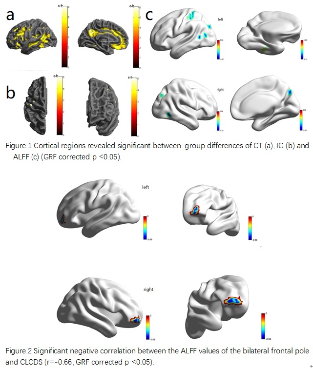
Figure.1 Cortical regions revealed significant between-group differences of CT (a), IG (b) and ALFF (c) (GRF corrected p <0.05)
Fig.2 Significant negative correlation between the ALFF values of the bilateral frontal pole and CLCDS (r=-0.66, GRF corrected p <0.05)