2607
Investigation of hypoxia after brain injury using a hypoxia-binding T1 contrast agent GdDO3NI1School of Biological and Health Systems Engineering, Arizona State University, Tempe, AZ, United States
Synopsis
In this study we have used the hypoxia-targeted MR contrast agent GdDO3NI, (a nitroimidazole-based T1 MRI contrast agent) to image the development of hypoxia in the rodent brain after traumatic brain injury (TBI). Our results indicate a statistically significant ~ 50% signal enhancement over baseline in the injury region using GdDO3NI compared to baseline values (~ 0%) observed with non-specific Gadoteridol (as control) at 3hours post injection. This study further demonstrates the utility of GdDO3NI in imaging tissue hypoxia and applicability to traumatic brain injury.
Introduction
An estimated 1.7 million TBI occur
annually in the U.S. accounting for over 50,000 deaths1. The pathology of TBI occurs from both immediate and delayed mechanisms
such as edema, ischemia, hemorrhage, finally resulting in highly heterogeneous
tissue damage2. Specifically, early posttraumatic hypoxia plays a significant role in
patient’s outcome3. Studies have reported about 30-50% of TBI patients in traumatic coma
have hypoxia upon arrival at an emergency room and the time to resuscitation
significantly affects the outcome4. Current diagnosis for TBI lacks the sensitivity to detect events such
as hypoxia that could provide information for better therapeutic intervention
and management. Magnetic resonance imaging (MRI) is a non-invasive imaging
modality with high resolution and myriad of information about anatomy and pathology
evolution of injury and therapeutic methods.
Materials and Methods
The target contrast agents, GdDO3NI, had been synthesized as described previously5,6. Gadoteridol was used as control, non-specific contrast agent. All animal studies were approved by Arizona State University’s Institute of Animal Care and Use Committee (IACUC) and were performed in accordance with the relevant guidelines. Traumatic brain injury was modeled using the well-established controlled cortical impact (CCI) injury model7 in mice brains. MRI studies were performed on a 7 T Bruker with surface coil. Mice were placed into the magnet right after the injury and pre-injection anatomical and T1 wt 3D gradient echo scans (2 cmX2 cmX 2cm, 128X64X64) were acquired before and after injection (every 10 min). Injection of the contrast agents was performed 1 hour after injury and follow up imaging started right after the injection.Results and Discussion
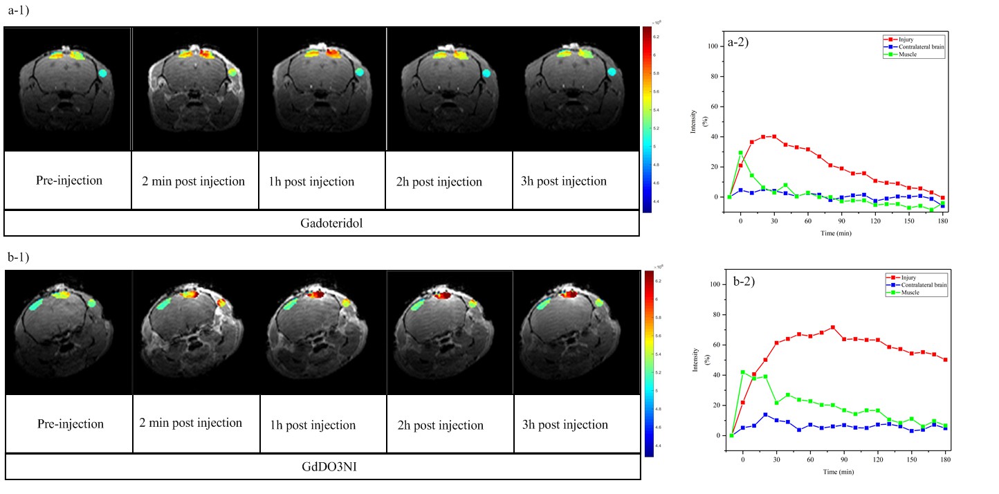
Fig. 1 shows the results of the two contrast agents on three different ROIs: injury, contralateral brain, and muscle on a representative animal. ROIs analysis in Matlab were performed by measuring the percentage enhancement of intensity in T1 weighted images acquired over three hours compared to the pre injection image. Contrast kinetics for gadoteridol show a similar trend of sharp increase and slower decrease of signal intensity for muscle and injured area. The washout of signal intensity in muscle is faster, changes in injured area are more slow and prolonged, which can be ascribe to the heterogenous nature of vascular leakage brain injury comparing to intact muscle. On the other hand, GdDO3NI delineates a different but interesting trend for muscle and injured brain. Once GdDO3NI was injected to blood stream, it washes out of the muscle region with similar kinetics as gadoteridol since there is no hypoxia; however, it stays in the injured region for significantly longer time, due to reduction and subsequent binding to proteins in the hypoxic regions. Fig 2 demonstrates 3D distribution of contrast enhancement in the injury regions for the two agents at 3hr post injection (2 a and b). Statistical comparisons (Fig 2c) that GdDO3NI shows significantly higher percentage enhancement (p<0.05) after 2 and 3 hours of injection compared to gatoteridol. IHC staining on sections are shown in Fig. 3 DAPI staining was visualized in blue and pimonidazole staining in green. Thus both cohorts (gadoteridol and GdDO3NI) show similar levels of hypoxia and contrast retention is only seen for GdDO3NI.Conclusion
The results clearly support that MRI, as a non-invasive imaging modality, with GdDO3NI provides important information regarding hypoxic regions in TBI. The MR results for hypoxia were validated by the gold standard method of IHC staining for pimonidazole, and showed an identical agreement to IHC outcomes.Acknowledgements
No acknowledgement found.References
1.Langlois JA, Rutland-Brown W, Wald MM. The Epidemiology and Impact of Traumatic Brain Injury: A Brief Overview. The Journal of Head Trauma Rehabilitation. 2006;21(5):375-378.
2.Alves JL. Blood–brain barrier and traumatic brain injury. Journal of Neuroscience Research. 2014;92(2):141-147.
3.Yan Edwin B. SL, Paul Eldho, Bye Nicole, Nguyen Phuong, Agyapomaa Doreen, Kossmann Thomas, Rosenfeld Jeffrey V., and Morganti-Kossmann Maria Cristina. . Post-Traumatic Hypoxia Is Associated with Prolonged Cerebral Cytokine Production, Higher Serum Biomarker Levels, and Poor Outcome in Patients with Severe Traumatic Brain Injury Journal of Neurotrauma. March 2014( 31(7)):618-629.
4.Ishige N, Pitts LH, Berry I, et al. The Effect of Hypoxia on Traumatic Head Injury in Rats: Alterations in Neurologic Function, Brain Edema, and Cerebral Blood Flow. Journal of Cerebral Blood Flow & Metabolism. 1987;7(6):759-767.
5.Rojas-Quijano FA, Tircsó G, Tircsóné Benyó E, et al. Synthesis and Characterization of a Hypoxia-Sensitive MRI Probe. Chemistry – A European Journal. 2012;18(31):9669-9676.
6.Gulaka PK, Rojas-Quijano F, Kovacs Z, Mason RP, Sherry AD, Kodibagkar VD. GdDO3NI, a nitroimidazole-based T 1 MRI contrast agent for imaging tumor hypoxia in vivo. JBIC Journal of Biological Inorganic Chemistry. 2014;19(2):271-279.
7.DOUGLAS H. SMITH HDS, JEAN S. PIERCE, KEVIN G. PERLMAN, KATHRYN E. SAATMAN, DAVID F. MEANEY, C. EDWARD DIXON, and TRACY K. McINTOSH. A Model of Parasagittal Controlled Cortical Impact in the Mouse: Cognitive and Histopathologic Effects Journal of Neurotrauma. January 2009(12(2)):169-178.
Figures