2605
MRI Detection of Amyloid Related Imaging Abnormalities (ARIA) in a Non-Human Primate Model of Sporadic Cerebral Amyloid Angiography at 7-Tesla1Department of Radiology, Bernard & Irene Schwartz Center for Biomedical Imaging & Center for Advanced Imaging Innovation & Research (CAI2R), New York, NY, United States, 2Preclinical Imaging Laboratory, Division of Advanced Research Technologies NYU Langone Health & NYU School of Medicine, New York, NY, United States, 3University of Texas MD Anderson Cancer Center, Houston, TX, United States, 4The University of Texas MD Anderson Cancer Center, Houston, TX, United States
Synopsis
Here we describe a non-invasive brain imaging method studying the pathogenesis and long-term effects of ARIA (amyloid-related imaging abnormalities) in an aged squirrel monkey (Saimiri Boliviensis), a non-human primate model of naturally occurring cerebral amyloid angiopathy. We investigated both ARIA-E, characterized by vasogenic edema, and ARIA-H, characterized by MRI evidence of hemosiderin deposits as potential biomarkers to use in a MRI methodology to monitor newly developed AD treatments.
Introduction
Early diagnosis of Alzheimer’s disease (AD), the most common form of dementia in the elderly, is critical in the management of patients with Aβ deposition and tauopathy/neurodegeneration occurring 10-15 years before cognitive decline.1 The use of biomarker imaging allows for the detection of AD pathologies with magnetic resonance imaging (MRI) as an important non-invasive diagnostic tool. Two kinds of amyloid related imaging abnormalities have been classified in mice and in humans: ARIA-E characterized by MRI evidence of vasogenic edema and/or effusion, and ARIA-H, characterized by MRI evidence of hemosiderin deposits.2 The pathobiology of ARIA remains poorly understood, in part due to the absence of an animal model of the disorder that would enable a contemporaneous analysis of tissue integrity in the affected region. The potential translatability to humans is enhanced by testing a non-invasive MRI protocol in non-human primates, which are a more biologically proximate AD model compared to transgenic mice. Here, we describe a brain imaging method studying the pathogenesis and long-term effects of ARIA in an aged squirrel monkey (Saimiri Boliviensis), a non-human primate model of naturally occurring cerebral amyloid angiopathy. Additional focus is to validate our novel targeted contrast agent, bi-functional ultrasmall superparamagnetic iron oxide nanoparticles coupled to polyethylene glycol (PEG) and Aβ peptide (USPIO-PEG-Aβ), in order to visualize amyloid plaque depositions in an aged squirrel monkey brain using a 3D multi gradient echo (MGE) sequence.Material and Methods
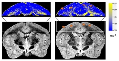
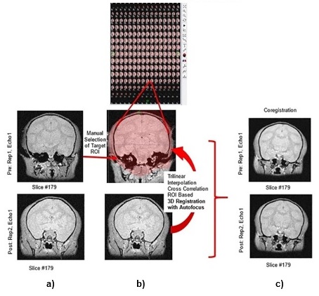
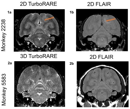
All MRI images were acquired on a Bruker Biospec 70/30 7-Tesla magnet equipped with actively shielded gradient coils and a Bruker commercially developed 86mm volume RF coil. 3D isotropic multi gradient echo images were acquired on one young and three aged monkeys, taken pre- and post-USPIO-PEG-Aβ injection, at various time points to further examine the pharmacokinetics of the nanoparticles. Each monkey was administered 3.5 mg of targeted nanoparticles per kg of body weight (within range prescribed in human studies) using a computer controlled syringe pump. To visualize edema burden, 2D Turbo RARE and FLAIR images were also taken and compared between one young and one aged monkey. The MGE images were processed by parametric map tool to yield R2* maps and coregistered using an ROI based rigid transformation in Firevoxel, an NYU software.3 Amyloid burden was performed by measuring R2* maps in various brain regions while edema was evaluated by comparing the brain bilateral symmetry using each brain as its own control. This was performed by both 2D TurboRARE and FLAIR sequence.Results and Discussion
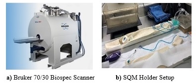
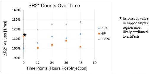
Figure 1 depicts the experimental setup to examine the non-human primates including the 7-Tesla Biospec 70/30 and the homemade setup to secure each subject examined in a reproducible manner. Figure 2 shows the R2* maps to evaluate the amyloid burden and Figure 3 the co-registration needed to compare between sessions and subjects thanks to the isotropic resolution. While additional quantitative approaches of amyloidosis are needed, we were able to use R2* maps as a surrogate marker of amyloid burden using USPIO-PEG-Aβ as an MRI probe as previously shown.4 Figure 4 summarizes the R2* difference between the various subjects in comparison to image sets prior to the injection. Figure 5 depicts the presence of edema by comparing the bilateral asymmetry where hyperintense regions can be seen in both 2D TurboRARE and FLAIR sequence in the aged monkeys.Conclusion
This MRI methodology is a potential non-invasive tool to monitor newly developed AD treatments to determine side effects and ability to reduce plaque burden. Future endeavors also include the confirmation of the presence of plaques and/or vasogenic edema with histology and the determination of the relationship between plaque burden and vasogenic edema. This approach suggests that diagnostic MRI methods to test the safety and efficacy of emerging therapies for AD may ultimately be feasible and translatable clinically.Acknowledgements
This work was supported, in part, by the NIH/XXX-1R01XXXXX (Scholtzova); NIH/XXX-R01XXXXX (Wisniewski); AHAF-ADR-A2008-155 (Wadghiri); Alz. Ass-IIRG-08-91618 (Wadghiri); NSF-DMR-1728858 (Montclare/Bonneau/Wadghiri;); NIH-1R01NS091552-01A1 (Bogdanov/Wadghiri); and was also performed at the Preclinical Imaging Laboratory, a shared resource partially supported by the NIH/SIG 1S10OD018337-01, the Laura and Isaac Perlmutter Cancer Center Support Grant NIH/NCI 5P30CA016087 and the NIBIB Technology Resource Center Grant NIH P41EB017183.References
1https://www.alz.org/alzheimers-dementia/research_progress/earlier-diagnosis
2Sperling RA, Jack CR, Black SE, et al. Amyloid-related imaging abnormalities in amyloid-modifying therapeutic trials: recommendations from the Alzheimer's Association Research Roundtable Workgroup. Alzheimers Dement. 2011;7(4):367-85.
3https://wp.nyu.edu/firevoxel/4Yang J, Wadghiri YZ, Hoang DM, et al. Detection of amyloid plaques targeted by USPIO-Aβ1-42 in Alzheimer's disease transgenic mice using magnetic resonance microimaging. Neuroimage. 2011;55(4):1600-9.
Figures