2600
Increased Intracortical R1 in the Motor Cortex of Exercising Older Adults1Neurology and Neurosurgery, McGill University, Montreal, QC, Canada, 2Neuroscience, McMaster University, Hamilton, ON, Canada, 3Brain Imaging Center Frankfurt/M., Frankfurt, Germany, 4Department of Sports Medicine, Institute of Sports Sciences, Goethe University, Frankfurt, Germany, 5Institute of Neuroradiology, Goethe University Hospital Frankfurt, Frankfurt, Germany, 6Department of Psychiatry and Psychotherapy, Charité University Medicine Berlin, Berlin, Germany, 7Institute of General Practice, Goethe University, Frankfurt, Germany, 8Department of Psychiatry, Psychosomatic Medicine and Psychotherapy, University Hospital Frankfurt, Frankfurt, Germany, 9Department of Psychology, Neuroscience and Behaviour, McMaster University, Hamilton, ON, Canada
Synopsis
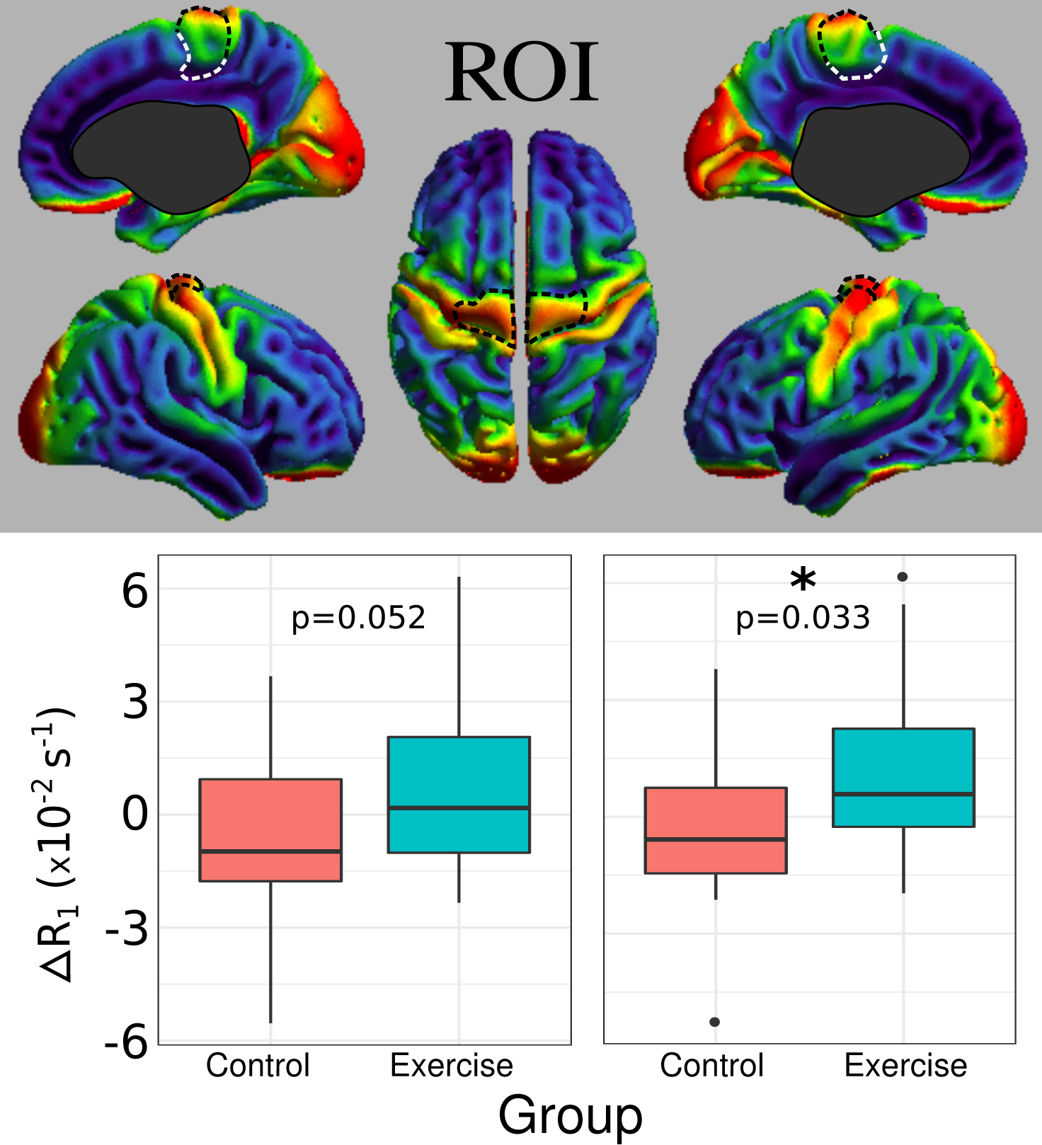
Exercise is known to be beneficial for brain health and performance; however, it is not known if changes in cortical microstructure underlie this effect. To investigate this, R1 maps acquired on cognitively healthy older adults (n=24, 65-90 years old) were analyzed before and after a 12-week exercise intervention. R1 prolongation indicating increased myelin levels were significant in the right (p=0.033) and trending in the left (p=0.052) leg motor regions with respect to a control group (n=22). ΔR1 correlated with aerobic cycling performance improvements (left: p=0.012, right: p=0.011). This study demonstrates that exercise promotes myelination in cortical motor regions.
Introduction
There is great interest in how exercise benefits the brain, with studies in humans demonstrating that exercise improves cognitive performance in people of all ages across a multitude of domains (reviewed in (1,2)), and also slows down age-related neuroanatomical degeneration (3-5). As of yet, the precise mechanisms underlying these findings are not fully understood. Thus, this study examined R1 maps reflecting myelination in the cortex to investigate if aerobic exercise in older adults can counteract age-related myelin degeneration and increase intracortical myelin levels.Methods
46 cognitively healthy participants aged 65-90 were randomly assigned to an exercise or control group. 24 participants completed the exercise intervention that consisted of 12 weeks of supervised cycle ergometer training (Optibike Med, Ergoline, Bitz, Germany), with three 30-minute sessions per week. 22 participants in the control group maintained their usual physical activity over the 12 weeks. All participants were imaged at weeks 1 and 13. Exercise intensity was set at each subject’s resistance in watts (W) at first ventilatory threshold (VT1) (equivalent to 64 ± 9% VO2max).
Images were collected on a 3T Siemens Magnetom Trio scanner (Siemens Medical, Erlangen, Germany). Quantitative mapping of T1 and proton density (PD) was conducted using the variable flip angle method with the following parameters: excitation angles 4°/24°, TR = 16.4 ms, TE = 6.7 ms, matrix size 256 × 224 × 160, isotropic spatial resolution 1 mm(6). Synthetic MPRAGE images with intrinsic B1+ and B1- correction were calculated from the T1 and PD maps (7)for cortical segmentation. Furthermore, R1 maps were calculated from R1=1/T1 to investigate changes in myelin. The T1-weighted synthetic MPRAGE data were used for cortical segmentation to ensure compatibility with the respective image processing software.
The cerebral cortex was segmented using custom Matlab scripts (vR2015a, https://www.mathworks.com), with manual inspection and corrections to gross topological errors using ITK-SNAP (version 3.4, http://www.itksnap.org). The white matter surface was generated using the FANTASM algorithm (8). Middle depth surfaces were generated and registered to the ICBM-152 atlas using CBS High-Res Brain Processing Tools Version v3.0 (www.nitrc.org/projects/cbs-tools) plug-ins within the MIPAV (mipav.cit.nih.gov) and JIST framework (www.nitrc.org/projects/jist/). ROI parcellation of the cortex was performed using the MarsAtlas (9).
The effect of exercise on ΔR1 during the study period in the dorsal medial precentral gyrus for each hemisphere was assessed using a t-test. The correlation between ΔR1 and improvement in submaximal aerobic performance was tested using the following general linear model: ΔR1~ R1(baseline)+ ΔVT1 O2/kg.
Results
To investigate activity-dependent myelination in exercise, an ROI analysis of the dorsal medial precentral gyrus was conducted. We found a significant R1 increase on the right side (p=0.033) and a trend for increased R1 on the left (p=0.052) (Figure 1).
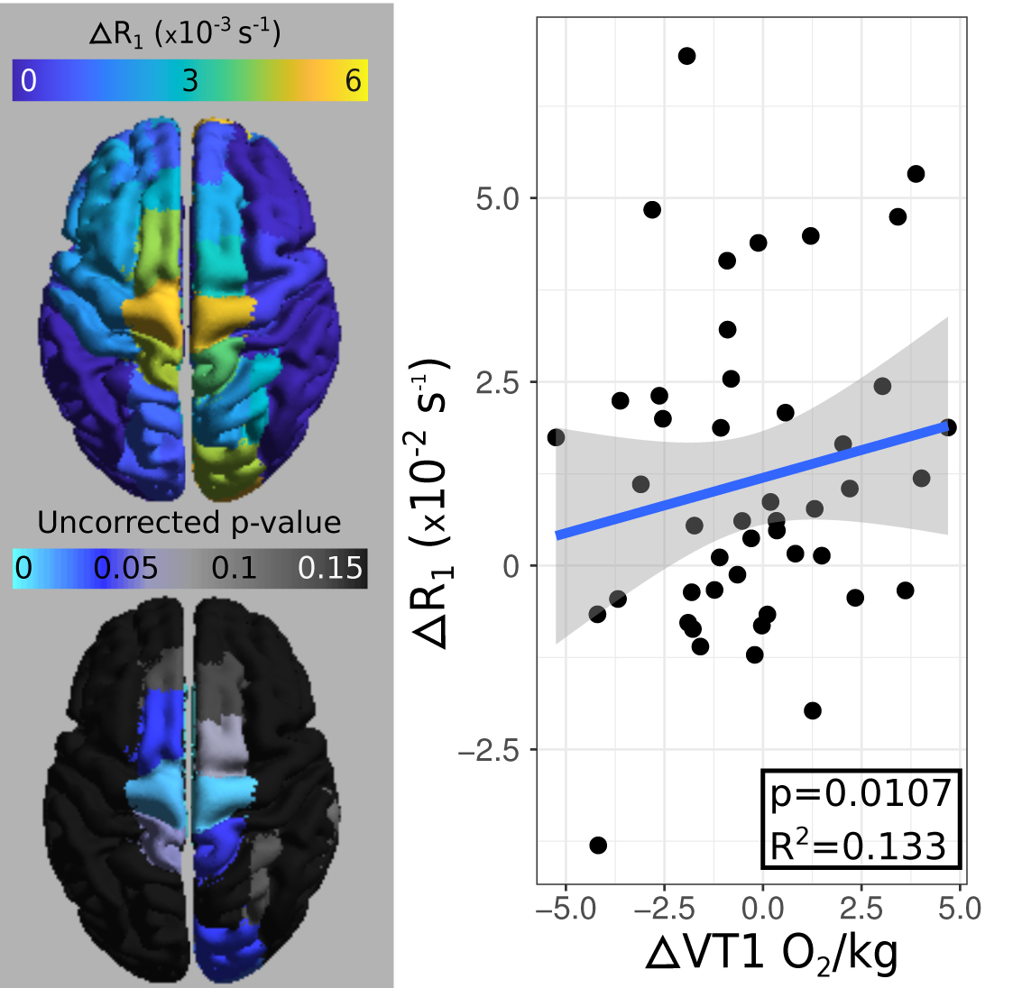
Subsequent exploratory analysis showed that R1 changes with ΔVT1 O2/kg were primarily found in the dorsal medial precentral gyrus on both hemispheres as evidenced by the elevated coefficients and decreased p-values from the linear models (Figure 2). Significant correlations between ΔR1 and ΔVT1 O2/kg were found in the left (p = 0.013) and right (p = 0.0107) dorsal medial precentral gyri. The scatterplot generated in Figure 2 for data from the right dorsal medial precentral gyrus allows for the visualization of the correlation between VT1 O2/kg metric, with the microstructural R1 metric.
Discussion and Conclusion
The a priori hypothesis driven analysis of ΔR1 in an ROI representing the leg representation in the primary motor cortex suggests that exercise increases intracortical myelin in this region. This finding reconfirms the results from experiments on mice which showed that myelination is an activity driven process (10). Furthermore, our exploratory analysis suggests that changes in R1 are positively correlated with improvements in exercise performance. Taken together these results demonstrate that the microstructure of the cortex can be modulated by external stimuli in old age to improve task performance.
Future work should aim to determine if exercise induced improvements in intracortical myelin extend beyond the motor cortex. Moreover, we need a better understanding if and how increased intracortical myelin also leads to improvements in other domains such as cognition. Revealing the mechanisms that underlie the beneficial effects of exercise on the brain could help to design interventions that might counteract age associated cognitive decline.
Acknowledgements
Parts of this study constitute the Master Thesis (M.A.) of Jonas Newlry and Sabrina Weber and the Thesis for a Medical Doctorate of Natkay Rahi, Alexandra Fischer and Katharina Dietz. We especially recognize the assistance of Mrs. Bianca Lienerth, the medical radiological assistant at the Brain Imaging Center and Mrs. Romy Schild, the medical assistant at the Department of Sports Medicine. We want to thank Horst Michaelis, former Director at the Cronstetten-Haus for his patronage.The trial has been granted by the Else-Kröner-Fresenius-Foundation, the Cronstetten Foundation and the Familie Schambach Foundation, all of them German non-profit foundations guaranteeing independency of research.References
1. Voss MW, Nagamatsu LS, Liu-Ambrose T, Kramer AF. Exercise, brain, and cognition across the life span. J Appl Physiol 2011;111:1505–1513. doi: 10.1152/japplphysiol.00210.2011.2. Hötting K, Röder B. Beneficial effects of physical exercise on neuroplasticity and cognition. Neuroscience and Biobehavioral Reviews 2013;37:2243–2257. doi: 10.1016/j.neubiorev.2013.04.005.3. Colcombe SJ, Erickson KI, Scalf PE, Kim JS, Prakash R, McAuley E, Elavsky S, Marquez DX, Hu L, Kramer AF. Aerobic exercise training increases brain volume in aging humans. J. Gerontol. A Biol. Sci. Med. Sci. 2006;61:1166–1170.4. Colcombe SJ, Erickson KI, Raz N, Webb AG, Cohen NJ, McAuley E, Kramer AF. Aerobic fitness reduces brain tissue loss in aging humans. J. Gerontol. A Biol. Sci. Med. Sci. 2003;58:176–180.5. Erickson KI, Voss MW, Prakash RS, et al. Exercise training increases size of hippocampus and improves memory. Proc. Natl. Acad. Sci. U.S.A. 2011;108:3017–3022. doi: 10.1073/pnas.1015950108.6. Volz S, Nöth U, Jurcoane A, Ziemann U, Hattingen E, Deichmann R. Quantitative proton density mapping: correcting the receiver sensitivity bias via pseudo proton densities. NeuroImage 2012;63:540–552. doi: 10.1016/j.neuroimage.2012.06.076.7. Nöth U, Hattingen E, Bähr O, Tichy J, Deichmann R. Improved visibility of brain tumors in synthetic MP-RAGE anatomies with pure T1 weighting. NMR Biomed. 2015;28:818–830. doi: 10.1002/nbm.3324.8. Pham DL. Robust fuzzy segmentation of magnetic resonance images. Computer-Based Medical Systems 2001.9. Auzias G, Coulon O, Brovelli A. MarsAtlas: A cortical parcellation atlas for functional mapping. Hum. Brain Mapp. 2016;37:1573–1592. doi: 10.1002/hbm.23121.10. Gibson EM, Purger D, Mount CW, et al. Neuronal activity promotes oligodendrogenesis and adaptive myelination in the mammalian brain. Science 2014;344:1252304–1252304. doi: 10.1126/science.1252304.Figures