2599
Investigation of brain plasticity during prolonged Braille learning in sighted subjects: a longitudinal diffusion MRI (dMRI) study1Laboratory of Brain Imaging, Neurobiology Center, Nencki Institute of Experimental Biology, Polish Academy of Sciences, Warsaw, Poland, 2Department of Psychology, Jagiellonian University, Cracow, Poland, 3Laboratory of Psychophysiology, Nencki Institute of Experimental Biology, Polish Academy of Sciences, Warsaw, Poland
Synopsis
Diffusion MRI can be used to evaluate the brain plasticity processes that occur during new skills acquisition. Commonly, one of the tasks used to investigate neuroplasticity of both blind and sighted subjects is Braille reading. In this work, we analyze DTI metrics based on
Introduction
Neuroimaging methods are sensitive to subtle changes of the brain tissue and demonstrate potential for human connectome understanding. dMRI could facilitate obtaining meticulous information related to the nature of training-induced plasticity processes in the brain. Brain plasticity is induced by a prolonged discrepancy between functional supply and environmental demands and is defined as the capability of the brain for reactive change in behavioral flexibility1. Based on MRI analysis it was proven that local neuroanatomical changes can be induced in adults while acquiring new skills2–5. Therefore, the purpose of the on-going study is to quantify to what extent plastic brain reorganization occurs in sighted subjects learning Braille reading, employing common characteristics of neural white matter development. In order to examine thoroughly the dynamics of the brain plasticity the key analyses were evaluated via dMRI biomarkers sensitive to white matter myelination6. Relatively little is understood about the dynamics of white matter reorganization. Thus, the proposed multiple time points study on sighted subjects who underwent Braille reading course introduces new opportunities for studying brain plasticity.
Methods
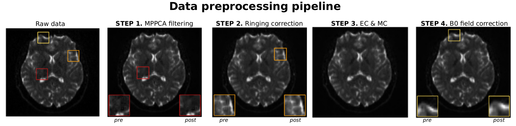
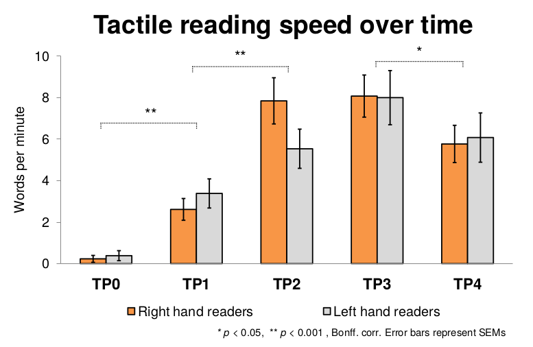
26 right–handed females (Age M = 23.2; SD = 2.1) underwent tactile Braille reading course lasting 8 months and covering 16 letters (A, B, C, D, E, I, K, L, Ł, M, O, P, S, T, U, Y). Half of the group declared preferences for reading with left hand after 3 months of training. Overall, 5 MRI sessions were conducted with 4 of them every 12 weeks (before the course - TP0, during tactile reading acquisition - TP1, TP2, TP3) and a follow-up 3 months after the course closure (TP4). dMRI was performed on 3T Siemens scanner with 12-channel coil using EPI pulse sequence with AP phase encoding and GRAPPA reconstruction. 64 diffusion sensitized for b-value b=1500 s/mm2 with 2 non-diffusion weighted images were acquired, comprising overall 80 axial slices of 128x128 base resolution. Additionally, field maps were collected to further correct the data for b0 field inhomogeneities. Data was denoised with Marchenko-Pasteur PCA filter7 (MRTrix), corrected for Gibbs ringing8 (MRTrix), motion and eddy current distortions9 (FSL eddy), and B0 field inhomogeneity10 (SPM Fieldmap toolbox). DTI metrics were computed with MRTrix software11. ANTs registration approach (buildtemplateparallel.sh) was run to create subject-specific templates based on 5 acquisitions. Next, mean subjects’ FA images were used to create study-specific group FA template with standard TBSS procedure12. In consequence, all subjects FA maps were registered to subject-specific and then group template using combined affine, rigid-body transformations and diffeomorphic warps. Normalized FA images were weightedly smoothed with [4,4,4] kernel within WM tissue probability mask segmented from group FA image13. Data was analyzed in within subjects ANOVA model in SPM12 at conservative FWE threshold p<0.05.Results
The key influence of preprocessing steps on the general quality of raw dMRI data is shown in Figure 1. Subjects successfully acquired tactile Braille skill, improving their reading speed over time. There was no effect of preferred hand, nor time by hand interaction (Figure 2).
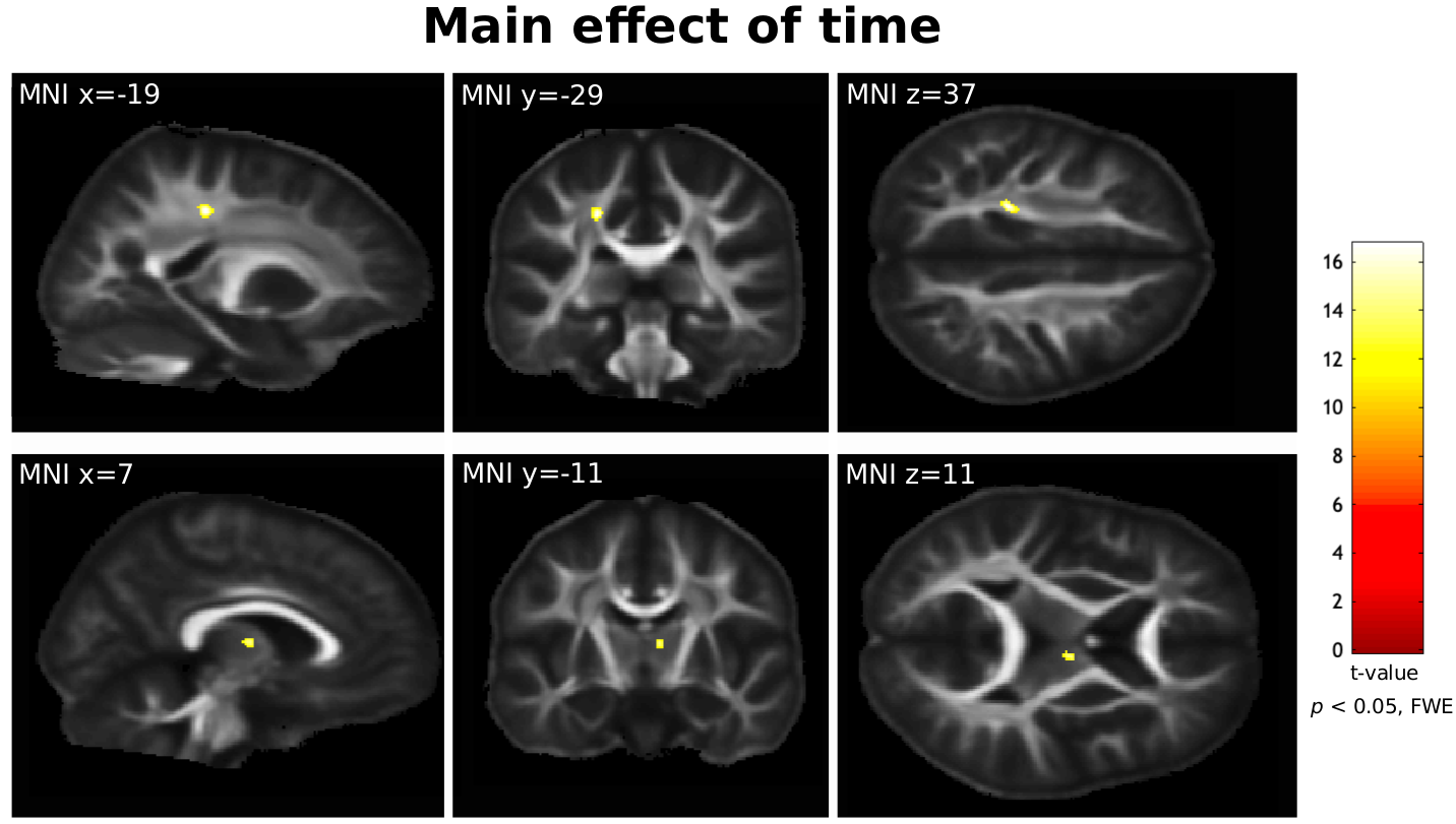
In voxel-wise analysis significant effect of time point showed that tactile reading training induced FA changes in various brain regions including left somatosensory, premotor, and motor regions as well as in surroundings of right Thalamus (Figure 3). Analysis showed that FA decrease detected in the thalamic area cover thalamocortical radiations that presumably connect thalamus with the cerebral cortex. For the case of somatosensory area reorganizations, the significant changes were observed mainly in posterior corona radiata and postcentral gyrus for the whole study group.
Conclusions
Our longitudinal study revealed dynamic brain reorganization due to tactile reading training in sighted subjects. Diffusion imaging methods show white matter reorganization associated with sensorimotor areas. Interestingly, the overall change in FA values within white-matter bundles may reflect strengthened anatomical connectivity within the sensorimotor network. We plan to further investigate the dynamics of these changes as well as compare results to control group in order to analyze the specificity of effects to tactile reading training.Acknowledgements
The study was financed by the National Science Center grant UMO-2014/14/M/HS6/00918.References
1. Lövdén, M., Bäckman, L., Lindenberger, U., Schaefer, S. & Schmiedek, F. A theoretical framework for the study of adult cognitive plasticity. Psychol. Bull. 136, 659–676 (2010).
2. Zatorre, R. J., Fields, R. D. & Johansen-Berg, H. Plasticity in gray and white: neuroimaging changes in brain structure during learning. Nat. Neurosci. 15, 528–536 (2012).
3. Engvig, A. et al. Memory training impacts short-term changes in aging white matter: a longitudinal diffusion tensor imaging study. Hum. Brain Mapp. 33, 2390–2406 (2012).
4. Bola, Ł. et al. Structural reorganization of the early visual cortex following Braille training in sighted adults. Sci. Rep. 7, 17448 (2017).
5. Draganski, B. et al. Changes in grey matter induced by training. Nature 427, 311–312 (2004).
6. Tardif, C. L. et al. Advanced MRI techniques to improve our understanding of experience-induced neuroplasticity. Neuroimage 131, 55–72 (2016).
7. Veraart, J., Fieremans, E. & Novikov, D. S. Diffusion MRI noise mapping using random matrix theory. Magn. Reson. Med. 76, 1582–1593 (2016).
8. Kellner, E., Dhital, B., Kiselev, V. G. & Reisert, M. Gibbs-ringing artifact removal based on local subvoxel-shifts. Magn. Reson. Med. 76, 1574–1581 (2016).
9. Andersson, J. L. R. & Sotiropoulos, S. N. An integrated approach to correction for off-resonance effects and subject movement in diffusion MR imaging. Neuroimage 125, 1063–1078 (2016).
10. Andersson, J. L., Hutton, C., Ashburner, J., Turner, R. & Friston, K. Modeling geometric deformations in EPI time series. Neuroimage 13, 903–919 (2001).
11. Veraart, J., Sijbers, J., Sunaert, S., Leemans, A. & Jeurissen, B. Weighted linear least squares estimation of diffusion MRI parameters: strengths, limitations, and pitfalls. Neuroimage 81, 335–346 (2013).
12. Smith, S. M. et al. Tract-based spatial statistics: voxelwise analysis of multi-subject diffusion data. Neuroimage 31, 1487–1505 (2006).
13. Lee, J. E. et al. A study of diffusion tensor imaging by tissue-specific, smoothing-compensated voxel-based analysis. Neuroimage 44, 870–883 (2009).
Figures