2593
Functional Brain Connectome and Mild Cognitive Impairment in Early Stage Parkinson Disease1Huaxi MR Research Center (HMRRC), Department of Radiology, West China Hospital of Sichuan University, Chengdu, China, 2Department of Neurology, West China Hospital of Sichuan University, Chengdu, China, 3Department of Radiology, Henan Provincial People’s Hospital & the People’s Hospital of Zhengzhou University, Zhengzhou, China
Synopsis
To use graph theory approaches and resting-state functional magnetic resonance imaging (MRI) to explore the brain functional network in patients with early stage Parkinson's disease (PD) and mild cognitive impairment (MCI). The whole-brain functional network was constructed by thresholding the Pearson correlation matrices of 90 brain regions. The results showed a less small-worldization characterized by decreased global integration and decreased local segregation in PD patients relative to healthy controls (HC). On the basis of these between-group difference in global and nodal properties, PD patients with MCI showed the lowest properties values, followed by PD patients with normal cognition and HC.
Introduction
Mild cognitive impairment (MCI) is a well-defined nonmotor manifestation of Parkinson's disease (PD), which greatly impairs function and quality of life and frequently progress to dementia.1 PD patients with MCI (PD-MCI) are candidates for disease-modifying intervention before irreversible changes occur, and this has promoted the research for objective imaging biomarkers to predict cognitive decline. Recent advances in psychoradiology, 2 particularly in conjunction with the graph theory analyses, allow the noninvasive characterization of brain network topologic organization in neuropsychiatric disease, including PD. However, little is known about how such changes might be associated with MCI. The main aim of this study was to explore functional brain network abnormalities related to cognition in patients with PD without dementia by using resting-state functional magnetic resonance imaging (MRI) and graph theory analyses.Methods
MRI scanning were carried out in Trio Tim (3T) MRI system (Siemens; Erlangen). Resting state functional MRI images were obtained from 39 early stage PD patients either with MCI (PD-MCI, N = 22) or with normal cognition (PD-NC, N =17), and 36 age- and gender-matched healthy controls (HC). Briefly, the whole brain was divided into 90 cortical and subcortical regions using the automated anatomic labeling atlas with each region representing a network node. Functional connectivity between these regions was established using Pearson correlations of the mean time series between all pairs of nodes.3 Whole-brain functional network was constructed by thresholding the resultant Pearson correlation matrix (90×90). Graph theory-based global (clustering coefficient Cp, characteristic path length Lp, normalized Cp γ, normalized Lp λ, local efficiency Elocal, global efficiency Eglobal, and small-worldness σ) and nodal (nodal degree, nodal efficiency, and nodal betweenness) network measures 4 were calculated, and then compared between PD and HC using nonparametric permutation tests. After significant between-group differences were identifed in the network metrics, we extracted the area under the curve values of topologic properties in each region that showed significant difference. Comparison among the PD-MCI, PD-NC and HC was performed by using analysis of variance followed by post hoc two-sample t tests. Finally, partial correlations were computed to examine relationships between these values and cognitive scores (attention and working memory, executive function, language, memory, and visuospatial function).5Results
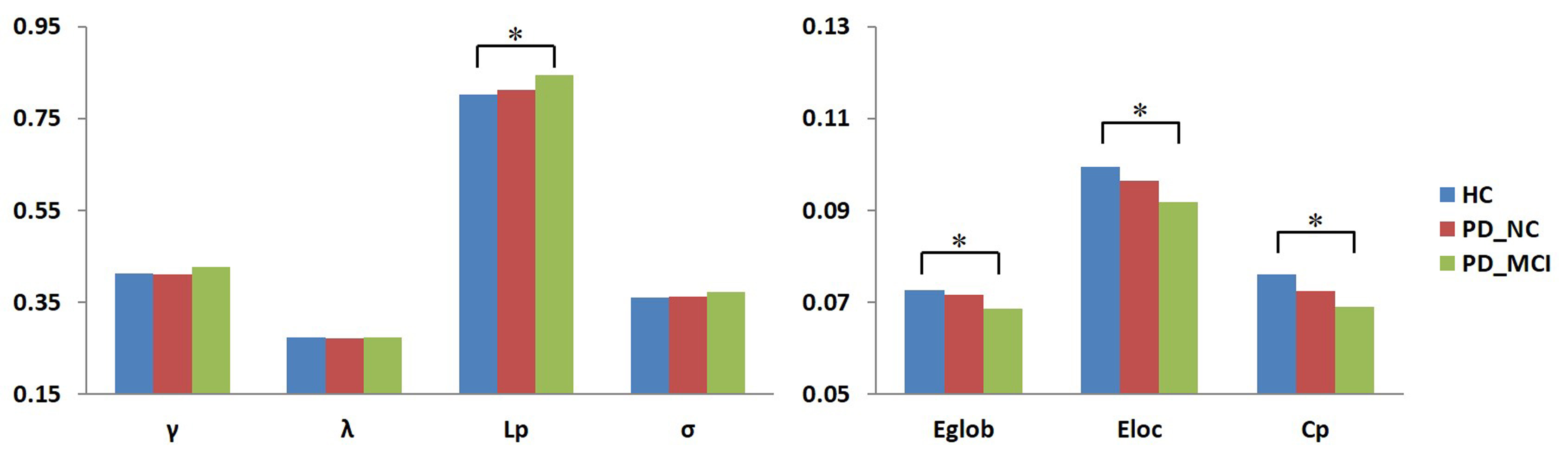
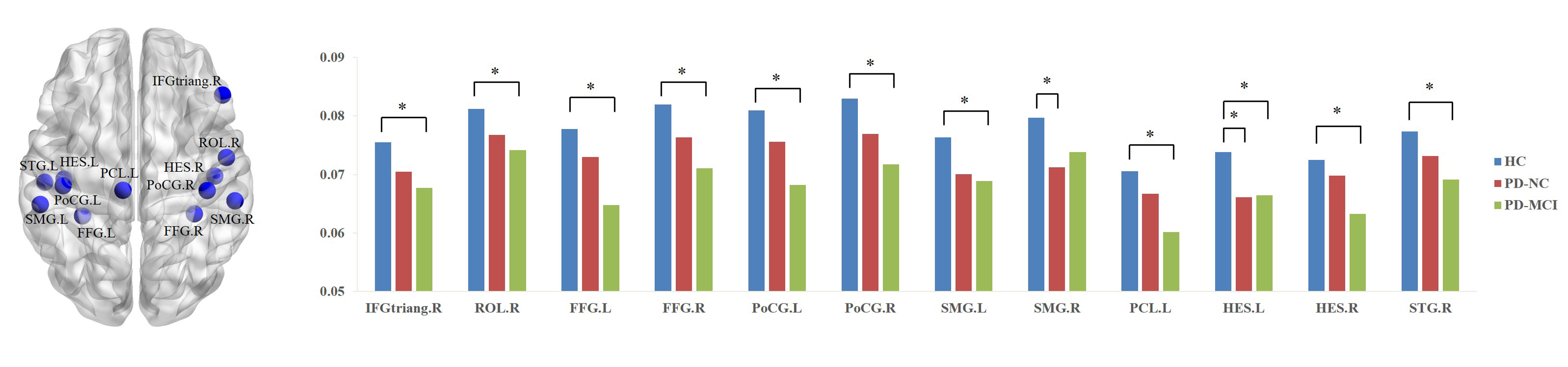
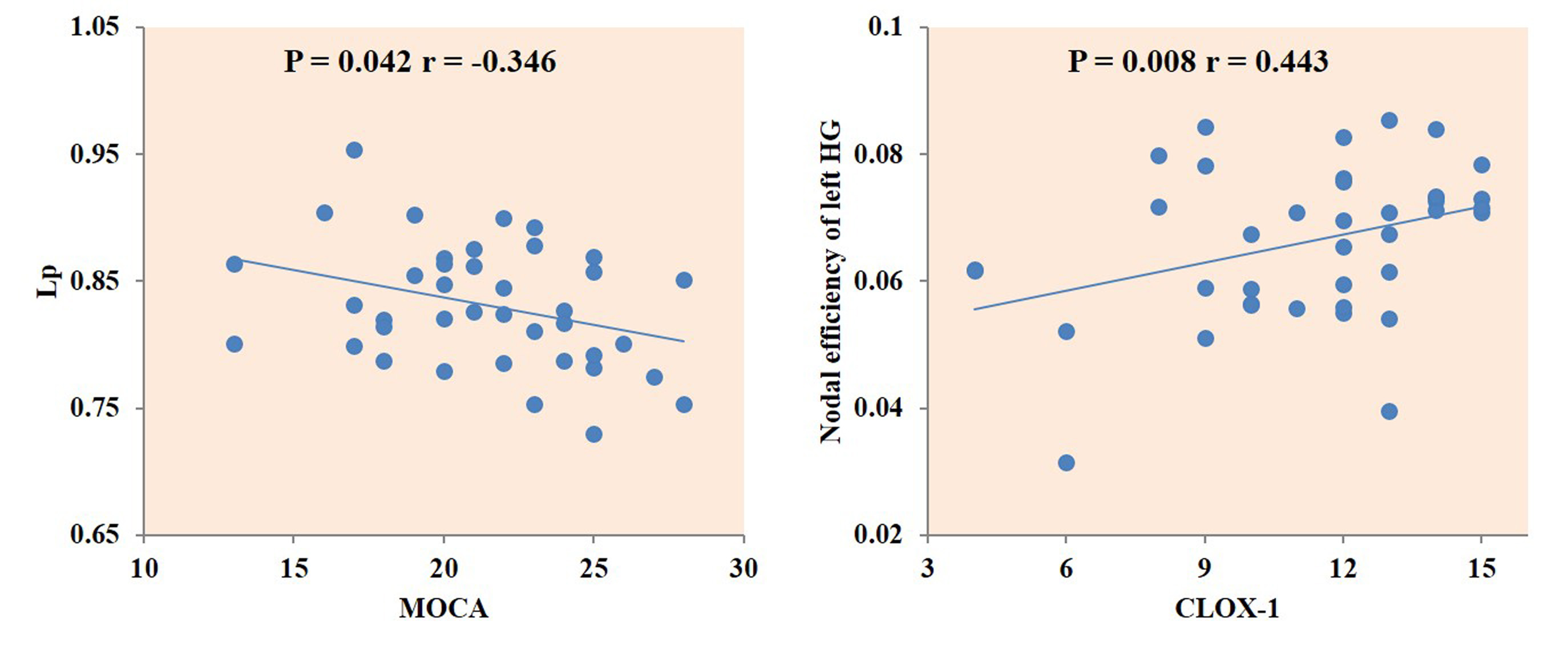
PD-MCI had lower Montreal Cognitive Assessment (MoCA) scores compared with PD-NC (19.1 vs. 24.3, p < .001). PD-MCI demonstrated significantly poorer performance on executive function, memory, and language abilities. Compared with HC, patients with PD showed a significant decrease in Cp, Elocal and Eglobal, and an increase in the Lp at the global level, and decreased nodal centralities in right inferior frontal gyrus, triangular part, right Rolandic operculum, bilateral fusiform gyrus, bilateral postcentral gyrus, bilateral supramarginal gyrus, left paracentral lobule, bilateral Heschl gyrus, and right superior temporal gyrus at the nodal level. In the majority of the global (Figure 1) and nodal properties (Figure 2), it was observed that PD-MCI group had the lowest properties values, while the properties values of PN-NC group was located in between HC and PD-MCI. Finally, MOCA score was negatively correlated with Lp, and the 10 points Clock Drawing Test (CLOX-1) score of executive function was positively correlated with nodal efficiency of left Heschl gyrus in the PD patients (Figure 3).Discussion
This study applied graph analysis combined with resting state functional MRI to assess large-scale brain functional networks in early stage PD patients. Compared with HC, the patients showed lower Cp, Eglobal and Elocal and longer Lp, indicating a shift toward 'less-small worldization' of the brain functional networks characterized by decreased local specialization and global integration. Patients with PD showed decreased nodal centralities in the default-mode network and sensorimotor cortex. Moreover, PD-MCI showed lowest properties values in the majority of these abnormalities compared to the other two groups. And these alterations were highly correlated with the cognitive scores implying that altered Lp and abnormal temporal regions are perhaps associated with pathogenesis of cognitive impairment in PD. These findings may provide insights into the neurobiology of PD and aid development of imaging biomarkers of cognitive decline.Conclusion
In this study, we tried to explore the MCI associated alterations on functional networks in PD patients. The results show that a clear pattern with lowest global and nodal properties values in PD-MCI, which are highly correlated with the global cognition and specific cognitive domain.Acknowledgements
This study was supported by the National Natural Science Foundation of China (Grant Nos. 81621003, 81761128023, 81220108013, 81227002, 81030027), the Program for Changjiang Scholars and Innovative Research Team in University (PCSIRT, grant IRT16R52) of China, the Changjiang Scholar Professorship Award (Award No. T2014190) of China, and the CMB Distinguished a Professorship Award (Award No. F510000/ G16916411) administered by the Institute of International Education.References
1. Pereira JB, Svenningsson P, Weintraub D, et al. Initial cognitive decline is associated with cortical thinning in early Parkinson disease. Neurology. 2014;82(22):2017-2025.
2. Lui S, Zhou XJ, Sweeney JA, et al. Psychoradiology: the frontier of neuroimaging in psychiatry. Radiology. 2016;281(2):357–372.
3. Suo X, Lei D, Li N, et al. Functional Brain Connectome and Its Relation to Hoehn and Yahr Stage in Parkinson Disease. Radiology. 2017;285(3):904-913.
4. Rubinov M, Sporns O. Complex network measures of brain connectivity: uses and interpretations. Neuroimage. 2010;52(3):1059–1069.
5. Litvan I, Goldman JG, Troster AI, et al. Diagnostic criteria for mild cognitive impairment in Parkinson's disease: Movement disorder society task force guidelines. Movement Disorders. 2012;27(3):349–356.
Figures