2591
Analysis of Structural Connectivity using Certain Nuclei as Seeds in Patients with Parkinson’s Disease1Center for Biomedical Imaging Research, Department of Biomedical Engineering, School of Medicine, Tsinghua University, Beijing, China, 2Wellcome Centre for Integrative Neuroimaging, FMRIB, Nuffield Department of Clinical Neurosciences, University of Oxford, Oxford, United Kingdom, 3Neuromodulation Center, Tsinghua University YuQuan Hospital, Beijing, China, 4School of Computer Science and Technology, Beijing Institute of Technology, Beijing, China
Synopsis
In this study, we aim to evaluate the relationships between fiber connectivity starting from specific nucleus regions to the whole brain and the Unified Parkinson’s Disease Rating Scale (UPDRS)-III scores in patients with Parkinson's disease. The results showed that the structural connectivity to the whole brain starting from bilateral internal global pallidus and caudate nucleus has significant negative correlations with the UPDRS-III scores. In the contrary, no significant correlations was found for the network starting from the putamen and external global pallidus. The strong negative correlation implies that these specific nuclei may play significant roles in the severity of Parkinson's disease. This finding is of great importance for further clinical research.
Introduction
Parkinson’s disease (PD) is one of the most common neurodegenerative diseases and is usually characterized by the asymmetric onset of motor symptoms. Deep brain stimulates (DBS) has been used to mitigate motor neurological symptoms by stimulating the subthalamic nucleus. How the stimulus affecting the brain structural connectivity is of importance to understand the underlying mechanism of this treatment. However, the networks starting from the likely involved nuclei still remain unclear. This study aimed to find the relationship between fiber connectivity starting from certain nuclei and disease progression in PD patients. Only nuclei investigated for PD previously, such as substantia nigra (SN), caudate nucleus (CN), global pallidus (GP), putamen (PUT), were tested here1. Diffusion tensor imaging (DTI) was used to map the topological organization of whole-brain starting from these nuclei. Unified Parkinson’s Disease Rating Scale (UPDRS)-III was used to measure the disease status.
Methods
1) Participants and data acquisition: 24 PD patients (age, 58.4 ± 9.6 years; male/female, 13/11) were recruited from Tsinghua University Yuquan Hospital, Beijing, China with informed consent. The UPDRS-III of each patient was scored by an experienced specialist. All scans were performed on a Philips 3.0T Achieva TX MR scanner (Philips Healthcare, Best, The Netherlands) using a 32-channel SENSE head coil. A single shot EPI DTI sequence was used with a multi-shell diffusion encoding scheme. The b-values were 0, 1000 and 2000 s/mm2. The imaging parameters were as follows: FOV = 224×224×140 mm3, resolution = 2.8×2.8×2.8 mm3, 50 slices in the whole brain, TE=85ms, TR = 5510 ms, flip angle = 90°.
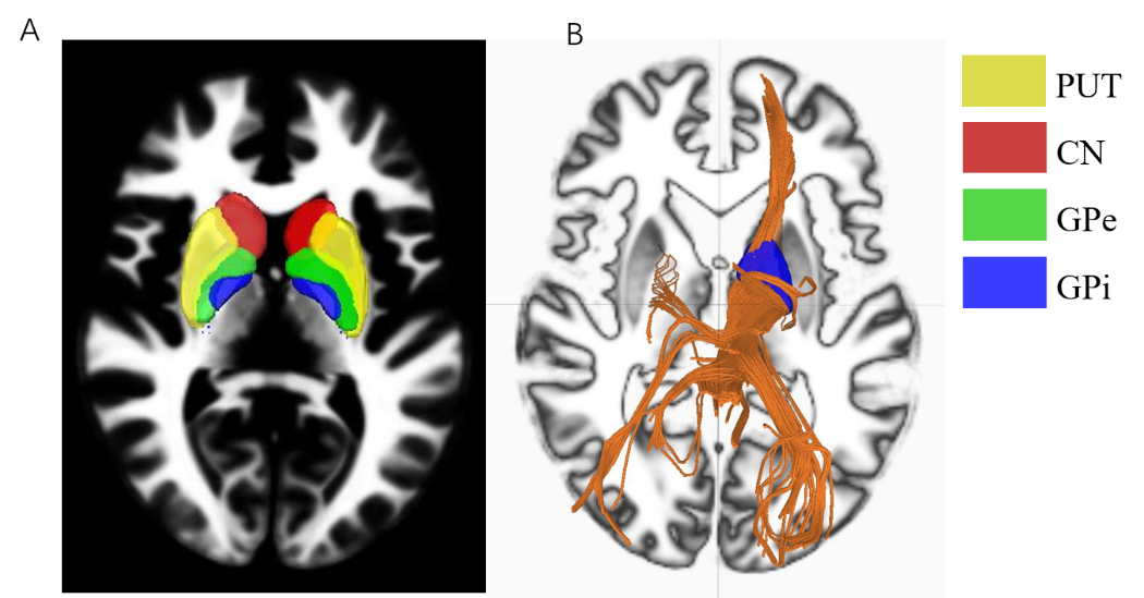
2) Image analysis: The diffusion images were analyzed in the Montreal Neurological Institute (MNI) template using q-space diffeomorphic reconstruction2 to obtain the spin distribution function3. A diffusion sampling length ratio of 1 was used, and the output resolution was 2 mm3. A deterministic fiber tracking algorithm was used. Seeding regions drawn manually were placed at bilateral CNs, GPes, GPis and PUTs as shown in Fig. 14. The angular threshold was set to 70 degrees. The step size was set to 1 mm. The anisotropy threshold was set to 0.4. The fiber trajectories were smoothed by averaging the propagation direction with 10% of previous directions. Tracks with length less than 20 mm were discarded. DSI Studio was used to obtain structural connectivity.
3) Statistical analysis: After the connectivity strength from the volumes of interest to whole brain was obtained, correlation analysis was performed to evaluate the relationships between the structural connectivity and UPDRS-III scores in these patients. Chi-Square test was applied to compare the obtained correlations.
Results and Discussion
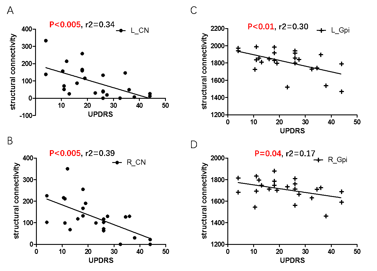
Patients with higher scores of UPDRS-III had lower reduction in structural connectivity in GPi and CN than those with lower scores (Fig. 2). However, in other nuclei regions including GPe and PUT, no significant correlations were observed (Fig. 3). GPi is the output nuclei of the basal ganglia and contains GABAergic neurons. By accepting movement requirement, it can disinhibit the thalamus and maintain the movement5. The negative correlation between GPi and UPDRS-III indicates the dysfunction of it. The low structural connectivity in GPi may affect its ability to receive inhibitory GABAergic signals, increasing the overall difficulty of starting and maintaining the movement. CN integrates spatial information with motor behavior movements and seems to link with spatially dependent motor preparation. Some literature also indicated that CN is involved in the recruitment of the motor system to support working memory6-7. The lack of structural connectivity leads to the decreasing ability to change behavioral choice based on target. The correlation is not obvious in GPe and PUT. This may indicate that their roles in the severity of PD are not important.Conclusion
In this study, we examined the microstructural deficits in PD patients through white matter integrity using DTI. Our data showed that the whole brain structural connectivity from the bilateral GPi and CN regions has significant correlations with the UPDRS-III scores. Patients with higher scores of UPDRS-III had lower reduction in structural connectivity than those with lower UPDRS-III scores. Our findings suggest that the microstructural deficits from GPi and CN might have an important role in the severity of PD.Acknowledgements
No acknowledgement found.References
[1] Conturo, et al. Tracking neuronal fiber pathways in the living human brain. PNAS. 1999; 96 (18) : 10422-10427
[2]Yeh et al. NTU-90: A high angular resolution brain atlas constructed by q-space diffeomorphic reconstruction. Neuroimage. 2011; 58(1):91-99.
[3] Yeh et al. Generalized q-Sampling Imaging. Medical Physics. 2010; 29(9):1626-1635.
[4] Naying He et al. Region-specific disturbed iron distribution in early idiopathic Parkinson's disease measured by quantitative susceptibility mapping. Human Brain Mapping. 2015; 36(11): 4407-4420.
[5] Morita et al. Distinct roles of the direct and indirect pathways in the basal ganglia circuit mechanism. Japanese Journal of Psychopharmacology. 2015;35 (5–6): 107–111.
[6] Grahn JA et al. The role of the basal ganglia in learning and memory: neuropsychological studies. Behavioural Brain Research. 2009;199(1): 53–60.
[7]Postle BR et al. Spatial working memory activity of the caudate nucleus is sensitive to frame of reference. Cognitive, Affective & Behavioral Neuroscience. 2003;3(2): 133–44.
Figures