2589
Atlas based Diffusion Abnormalities in Substantia Nigra in Parkinson's Disease1Symbiosis Centre for Medical Image Analysis, Symbiosis International University, Pune, India, 2Department of Neurology, National Institute of Mental Health and Neuro Sciences, Bangalore, India, 33Department of Clinical Neurosciences, National Institute of Mental Health and Neurosciences, Pune, India, 44Department of Neuroimaging & Interventional Radiology, National Institute of Mental Health and Neuro Sciences, Bangalore, India
Synopsis
Parkinson’s disease (PD) is characterized by neuronal loss of dopaminergic neurons in the substantia nigra (SN). This study aims to gain deeper insights into the abnormalities in SN by evaluating the diffusion metrics of the SN in a large cohort of patients with PD. To precisely delineate the SN, neuromelanin sensitive MRI images were obtained from a set of healthy controls and were used to create a probabilistic atlas of the SN. Using this atlas, we observed significantly higher radial and mean diffusivity of bilateral SN in patients with PD suggesting microstructural abnormalities that could potentially serve as bio-markers for PD.
Introduction
Parkinson’s disease (PD) is characterized by degeneration of dopaminergic neurons in the substantia nigra (SN). The role of routine neuroimaging in the diagnosis of PD, however, has been limited owing to the inability to adequately visualize and detect changes in the SN. A recently described neuromelanin sensitive sequence overcomes this limitation and has been used to differentiate between patients with PD and healthy control[1]. Existing studies have demonstrated volume loss as well as abnormalities in the diffusion tensor imaging (DTI) based measures of fractional anisotropy (FA) and increase in the mean diffusivity (MD) of the SN in patients with PD[2, 3]. Nonetheless, there is variability of results from these studies and this may be due to inconsistencies in the methods of delineating the SN. To mitigate this, our study aims to first create a probabilistic atlas of the SN using manually drawn regions of interest of the SN as visualized on the neuromelanin sensitive sequence for twenty eight healthy controls. We then employ this atlas to map the SN to a large population of PD patients to evaluate the diffusion metrics.Methods
Seventy-nine patients with PD and 37 age and gender matched healthy controls were scanned on a 3T Philips Achieva MRI scanner using a 32-channel head coil. Diffusion weighted images (DWI) for these subjects were acquired using a single-shot spin-echo EPI sequence: TR/TE =8583-9070ms/60-62ms; voxel-size=1.75x1.75x2mm; diffusion gradients=15 with b-value=1000s/mm2 and single b=0s/mm2 image. For creating a probabilistic atlas of SN, a different set of controls (n=28) were scanned on the same scanner. T1-weighted images was acquired using TR/TE=8.06/3.6ms, voxel-size=1x1x1mm, and neuromelanin contrast sensitive sequences i.e. the spectral presaturation with inversion recovery (SPIR) sequence was acquired using TR/TE: 26/2.2ms, field of view: 180x180x50mm; voxel size: 0.9x0.9x1mm; number of slices: 50. Bilateral substantia nigra ROIs (snROIs) were created by manual segmentation followed by registration to the T1 images using affine transformations and finally were transformed to the MNI space using ANTS[4]. A probabilistic atlas of SN in MNI space was created and thresholded at 0.5. For the PD subjects and the matched controls described above, DWI images were pre-processed using eddy correction, removing the non-brain tissue and fitting the tensor model to compute the diffusion measures of FA, MD, radial and axial diffusivity (RD, AD). FA maps of all subjects were registered to the FMRIB58 image using ANTS and the transformation matrix of this registration was used to register the remaining maps to MNI template. Diffusion measures were extracted for left and right SN for all subjects in the MNI space. Statistical analysis on mean FA, MD, RD and AD was performed using a MANCOVA model, with age and gender as covariates. The duration of illness (DoI), unified PD rating scale (UPDRS) and levodopa equivalent daily dosage (LEDD) of PD patients were correlated with the diffusion measures after correcting for age and gender.
Results
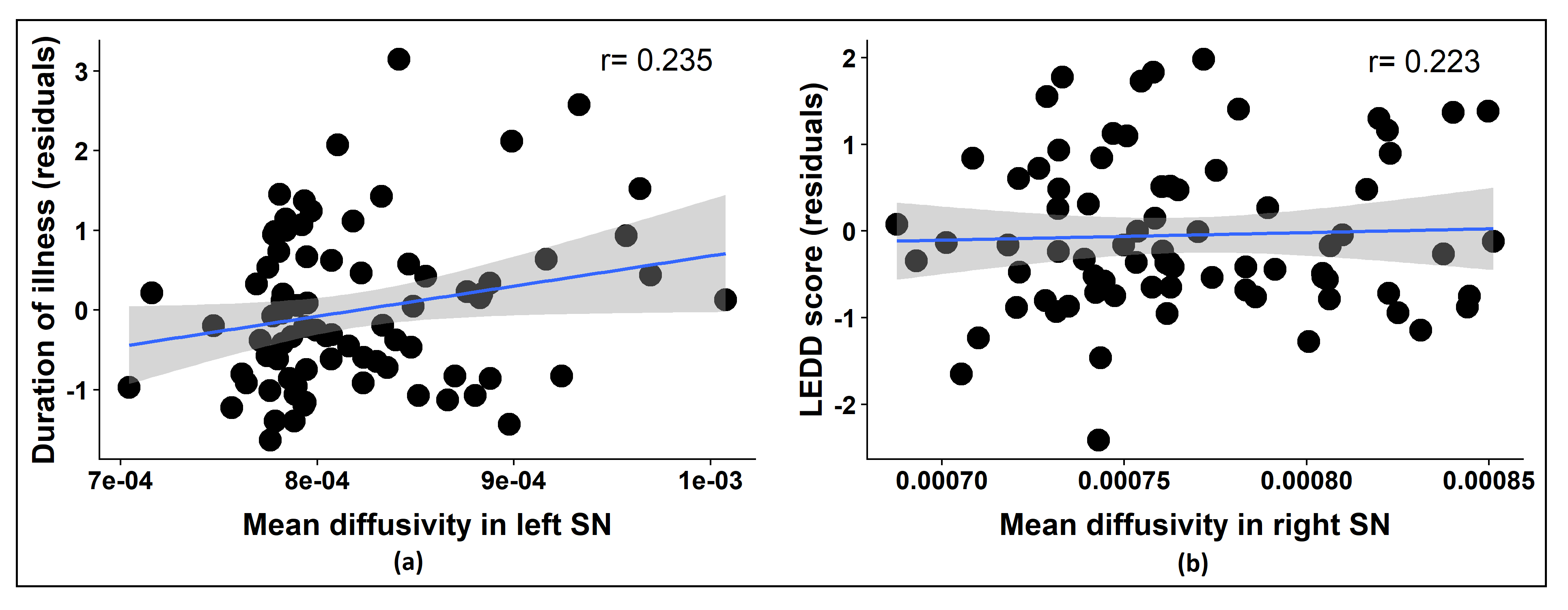
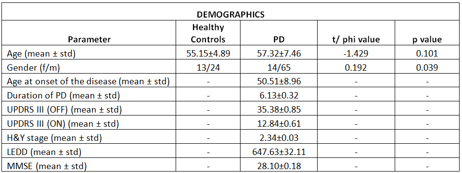
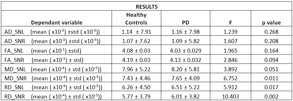
Table 1 shows the demographic information for the PD dataset. Figure 1 displays the probabilistic and thresholded (50%) atlas of the SN computed from the neuromelanin sensitive images of 28 controls. For our PD dataset, significant differences were observed in RD and MD values in the SN. Both the right and left SN had significantly higher RD and MD in patients with PD in comparison to controls (RD: right snROI: f=10.325, p<0.01, left snROI: f=5.904, p<0.05; MD: right snROI: f=3.925, p=0.05, left snROI: f=6.757, p<0.05). No significant differences were observed for FA and AD. Age significantly co-varied for all diffusion parameters except for right RD. Significant positive correlation was found between MD of left SN and DoI (r=0.235,p=0.037) and between MD of right SN and LEDD score (r=0.223,p=0.049).Discussion
In this work, we created a probabilistic atlas of the SN using snROIs derived from neuromelanin sensitive images, and applied this atlas to define the SN in patients with PD in order to accurately delineate the SN and evaluate the diffusion measures. On application of this atlas to a large population with PD, we observed significant abnormalities in RD and MD in PD in comparison to controls. Moreover, the MD measures correlated with the LEDD and DoI. In summary, our findings are suggestive of microstructural abnormalities of the SN in patients with PD and measurement of RD and MD of the SN can be established as a potential surrogate marker for diagnosis and monitoring of disease progression of PD.Acknowledgements
Department of Science and Technology –Science Education and Research Board (DST-SERB) (ECR/2016/000808) provided partial funding for setting up the computing facility.References
1. Prasad, S., et al., Three-dimensional neuromelanin-sensitive magnetic resonance imaging of the substantia nigra in Parkinson's disease. Eur J Neurol, 2018. 25(4): p. 680-686.
2. Aziz M, A.C., Castellanos G et al, Dynamic Atlas-based segmentation and quantification of Neuromelanin-rich brainstem structures in Parkinson disease. IEEE Trans Med Imaging, 2018.
3. Deng, X.Y., et al., A meta-analysis of diffusion tensor imaging of substantia nigra in patients with Parkinson's disease. Sci Rep, 2018. 8(1): p. 2941.
4 http://stnava.github.io/ANTs/.
Figures