2579
Longitudinal Changes in Cerebral Blood Flow Calculated by Arterial Spin Labeling MRI in Parkinson’s Disease1Biomedical Engineering Institute, Bogazici University, Istanbul, Turkey, 2Hulusi Behcet Life Sciences Research Center, Istanbul University, Istanbul, Turkey, 3Institute of Psychology and Cognition Research, University of Bremen, Bremen, Germany, 4Department of Neurology, Istanbul University, Istanbul, Turkey, 5Department of Physiology, Istanbul University, Istanbul, Turkey
Synopsis
The aim of this study is to monitor perfusion changes over one and a half years in Parkinson’s disease with mild cognitive impairment (PD-MCI). Cerebral blood flow (CBF) maps were created for baseline and follow-up scans by fitting a kinetic curve model for each pixel of arterial spin labeling MR images. The CBF maps were registered to MNI152 brain atlas, and perfusion changes were assessed at 119 distinct brain regions. The CBF of PD-MCI patients decreased at occipital fusiform gyrus, right occipital fusiform gyrus, anterior part of left supramarginal gyrus, and anterior part of right middle temporal gyrus over time.
Introduction
Mild cognitive impairment in Parkinson’s disease (PD-MCI) is an intermediate stage between cognitively normal PD (PD-CN) and dementia (PDD). PD-MCI patients have an increased risk of developing dementia.1 Previous studies have reported that abnormal perfusion patterns could provide information about the diagnosis of cognitive decline in PD.2,4 Arterial spin labeling MRI (ASL-MRI) provides information about perfusion parameters by measuring cerebral blood flow (CBF).5 The main aim of this study is to quantify the longitudinal change in perfusion measurements of PD-MCI patients.Methods
23 PD-MCI patients, who were previously diagnosed based on an extensive neuropsychological test battery, were included in this study. All participants were scanned on a 3T Philips MR scanner with a 32-channel head coil after they received their medications to reduce motion artifacts. ASL-MR images were obtained from six slices at eight different inversion times (TIs) by using STAR labeling with Look-Locker readout. For each slice, data acquisition was repeated 30 times to increase signal to noise (SNR). Also, acquisition was repeated three times to cover the whole brain. Follow-up analysis was carried out after one and a half years, and the patients were scanned by employing the same ASL-MRI sequence. A program was written in MATLAB R2018a (MathWorks Inc., Natick, MA) to calculate the CBF maps. The main magnetization, M0, was calculated for each pixel of control images by fitting multiple TI data to the longitudinal relaxation equation by taking into account the flip angle effect. CBF maps were calculated by using the combination of the model proposed by Gunther et al. 6 and the correction of arterial blood volume proposed by Chappell et al 7. CBF maps were registered into MNI152 brain atlas by using FMRIB Software Library (FSL) (http://fsl.fmrib.ox.ac.uk/fsl/fslwiki/). CBF values were estimated at 119 distinct brain regions defined by Harvard-Oxford cortical and subcortical structural atlases and the MNI structural atlas. A Wilcoxon signed-rank test was used to assess the CBF changes over time. A P-value of less than 0.05 was considered as statistically significant.Results
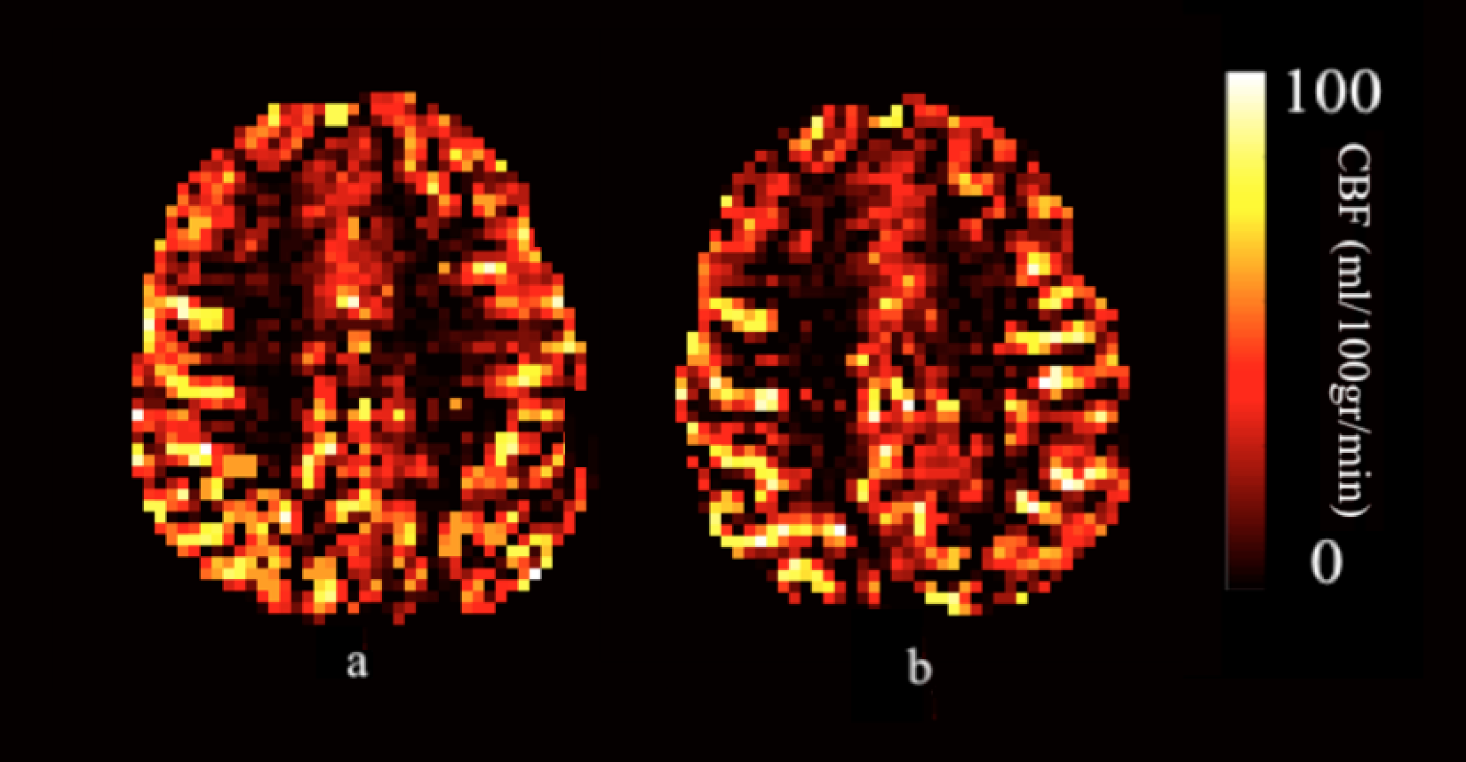
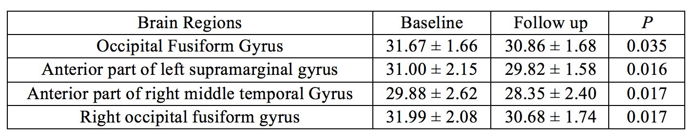
Figure 1 shows CBF maps calculated for one PD-MCI subject at baseline (a) and follow-up (b) time points. Based on neuropsychological test assessments, five PD-MCI patients converted from MCI to CN, while the status of 18 PD-MCI patients didn’t change. Table 1 shows the mean (+std) CBF values of 18 PD-MCI patients at the brain regions that had a change over time. At follow-up, PD-MCI patients had lower perfusion at occipital fusiform gyrus, right occipital fusiform gyrus, anterior part of left supramarginal gyrus and anterior part of right middle temporal gyrus compared to baseline measurements. There were not any statistically significant differences between baseline and follow-up CBF values of the patients who converted to CN.Discussion
In line with our study results, previous studies have reported hypoperfusion primarily at the parieto-occipital areas for PD.2-4 Right anterior temporal gyrus has been correlated with visual recognition scores.8 The supramarginal gyrus, which is a portion of the parietal lobe, has a role in visual word recognition.9 The progressive perfusion decrease might be related to visual deficits of PD.Conclusion
In conclusion, CBF values measured by ASL-MRI might help in monitoring the progression of PD-MCI, and might be useful for providing a biomarker for the cognitive assessment of Parkinson’s disease. In future, CBF values will be assessed at brain parcellations obtained from resting state fMRI.10 Partial volume correction will be applied in ASL data analysis to compare pure gray matter perfusion values of subjects.11Acknowledgements
This study was supported by TUBITAK project #115S219 and the Ministry of Development project #2010K120330.References
1. Poewe W, Gauthier S, Aarsland D, et al. Diagnosis and management of Parkinson's disease dementia. Int J Clin Pract. 2008; 62: 1581-7.
2. Chao LL, Buckley ST, Kornak J, et al. ASL perfusion MRI predicts cognitive decline and conversion from MCI to dementia. Alzheimer Dis Assoc Disord. 2010; 24: 19-27.
3. Melzer TR, Watts R, MacAskill MR, et al. Arterial spin labelling reveals an abnormal cerebral perfusion pattern in Parkinson's disease. Brain. 2011; 134: 845-55.
4. Le Heron CJ, Wright SL, Melzer TR, et al. Comparing cerebral perfusion in Alzheimer's disease and Parkinson's disease dementia: an ASL-MRI study. J Cereb Blood Flow Metab. 2014; 34: 964-70.
5. Petcharunpaisan S, Ramalho J and Castillo M. Arterial spin labeling in neuroimaging. World J Radiol. 2010; 2: 384-98.
6. Gunther M, Bock M and Schad LR. Arterial spin labeling in combination with a look-locker sampling strategy: inflow turbo-sampling EPI-FAIR (ITS-FAIR). Magn Reson Med. 2001; 46: 974-84.
7. Chappell MA, MacIntosh BJ, Donahue MJ, Gunther M, Jezzard P and Woolrich MW. Separation of macrovascular signal in multi-inversion time arterial spin labelling MRI. Magn Reson Med. 2010; 63: 1357-65.
8. Rice GE, Lambon Ralph MA and Hoffman P. The Roles of Left Versus Right Anterior Temporal Lobes in Conceptual Knowledge: An ALE Meta-analysis of 97 Functional Neuroimaging Studies. Cereb Cortex. 2015; 25: 4374-91.
9. Stoeckel C, Gough PM, Watkins KE and Devlin JT. Supramarginal gyrus involvement in visual word recognition. Cortex. 2009; 45: 1091-6.
10. Schaefer A, Kong R, Gordon EM, et al. Local-Global Parcellation of the Human Cerebral Cortex from Intrinsic Functional Connectivity MRI. Cereb Cortex. 2018; 28: 3095-114.
11. Asllani I, Borogovac A and Brown TR. Regression algorithm correcting for partial volume effects in arterial spin labeling MRI. Magn Reson Med. 2008; 60: 1362-71.
Figures