2577
Meta-analysis of the diagnostic effect size of neuromelanin MRI in Parkinson’s disease1Department of Radiological Sciences, University of Nottingham, Nottingham, United Kingdom
Synopsis
The clinical diagnosis and monitoring of Parkinson’s disease (PD) remain challenging which prompted substantial research efforts to develop pathophysiological meaningful biomarkers. Depigmentation of the substantia nigra (SN), pars compacta, is a pathological hallmark of PD that can be detected by neuromelanin-sensitive MRI (NM-MRI). We undertook a meta-analysis on the pooled diagnostic accuracy of NM-MRI in 14 case-control studies including 755 subjects (427 PD). We show a consistent decrease of SN NM signal in PD vs controls independent of the acquisition and analysis methods with a pooled standardized mean difference of SMD=1.06, 95% CI, 0.84, 1.28, p<0.00001. In conclusion, this meta-analysis supports NM-MRI metrics as a diagnostic biomarker of PD.
Introduction
Parkinson’s disease (PD) is the most common movement disorder and the second most common neurodegenerative disorder that affects human population. The diagnosis of PD remains challenging during the early symptomatic disease phase when already a substantial number of neurons in the substantia nigra (SN) have died1. A further challenge is the objective tracking of the disease. These unmet diagnostic needs prompted major research efforts by several groups to develop a pathophysiological meaningful biomarker that could be used as a diagnostic tool and to monitor the progression of PD. Recent studies suggest that magnetic resonance imaging made sensitive to neuromelanin (NM-MRI) is able to depict and quantify the depigmentation of NM loss in PD 2, 3, 4. Several studies reported clinically useful diagnostic accuracy using a wide range of scan protocols and metrics, but the consistency of findings needs to be investigated. This meta-analysis aimed to synthesise published evidence of NM-MRI to detect and quantify neuromelanin loss in SN as a step to qualify NM-MRI as a diagnostic marker in PD.Methods
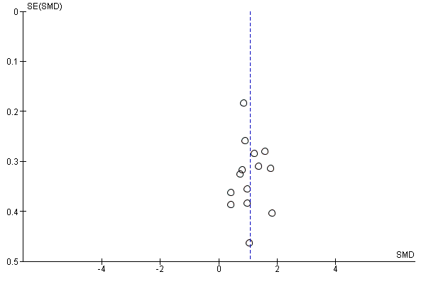
The meta-analysis was undertaken in accordance with the ‘preferred reporting items for systematic reviews and meta-analysis’ PRISMA statement5. Studies on NM-MRI were identified through a systematic online database, PubMed and Web of Science. Studies were selected based on predefined inclusion criteria, such as the subjects had to be scanned at 3T using dedicated neuromelanin sequences, the patients must be diagnosed using UK PD Brain Bank Criteria or Brain Clinical Diagnosis Criteria, the healthy control (HC) subjects are without movement disorder and at least 6 subjects included per group. We report standardized mean differences of NM related signal measurements within the SN to account for a range of different metrics used (e.g. contrast ratio, volume, width, areas etc.). Publication bias was assessed by a funnel plot.Results
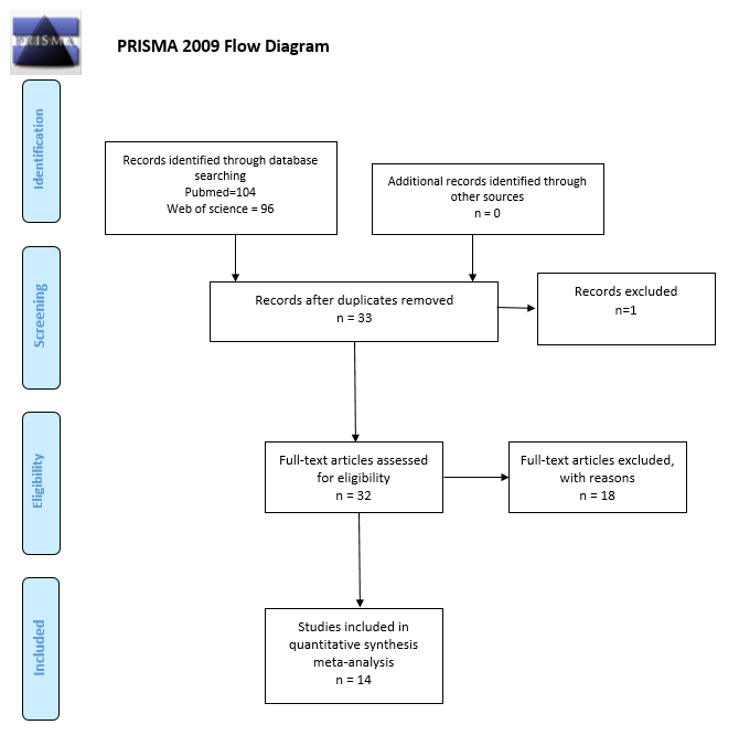
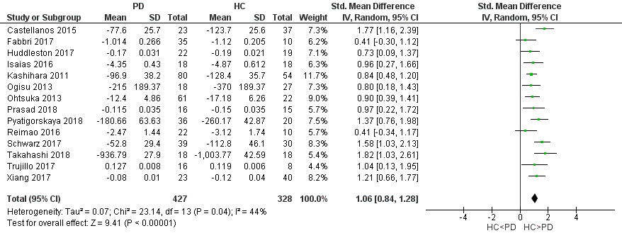
The literature search till 24th September 2018 resulted in 200 articles from which 14 articles were finally included for data extraction (Figure 1). This resulted in 755 subjects, 427 PD, 328 HC. The age range of the population was between 56 to 72 years and the patients’ disease was at an early stage (UPDRS and Hoehn and Yahr) for most of the studies. We found a consistent decrease of NM-related signal intensity in the SN of the subject is with PD vs. HC, which showed an overall large effect size (SMD=1.06, 95% CI, 0.84, 1.28, p<0.00001). However, there was a moderately large heterogeneity of study results (I2=44%, p=0.04) (Figure 2). The funnel plot illustrated no clear asymmetry in all studies thus not pointing to a major publication bias among those studies (Figure 3).Discussion
The meta-analysis of 14 case-control studies confirms that neuromelanin loss can be consistently detected in PD using different scanner platforms, acquisition and analysis protocols of NM-MRI (e.g. voxel intensity, area, volume, and contrast ratio). The pooled standardized mean difference of 1.06 suggests a large diagnostic effect size. This outcome is consistent with the pathological hallmark of SN depigmentation in PD that in turn is associated with postmortem confirmation of SN neuronal loss in PD6,7. The moderately high heterogeneity (I2=44%) may be due to inconsistencies in the MRI acquisition protocols, data analysis, and ROI selection but true biological heterogeneity cannot be excluded (e.g. different severity, stages of the disease, subtypes). Nevertheless, we recommend future research to further standardize NM-MRI to improve the consistency of the outcome prior to clinical practice. Our meta-analysis is limited by a lack of data to examine the diagnostic accuracy of the NM-MRI. Further, the small number of subjects in each study might induce bias and reduce the statistical power of this meta-analysis.Conclusion
In conclusion, this meta-analysis further supports NM-MRI as a promising diagnostic biomarker in PD. Further research is warranted to validate the diagnostic power in larger datasets ideally with a standardized signal acquisition as well as analysis protocols. Finally, the potential value of NM-MRI as a progression marker should be investigated.Acknowledgements
The first author is supported by the Ministry of Education Malaysia.References
1. Cheng, H. C., Ulane, C. M. & Burke, R. E. Clinical progression in Parkinson disease and the neurobiology of axons. Ann. Neurol. 67, 715–725 (2010).
2. Schwarz, S. T., Xing, Y., Tomar, P., Bajaj, N. & Auer, D. P. In vivo assessment of brainstem depigmentation in Parkinson disease: potential as a severity marker for multicenter studies. Radiology 283, 789–798 (2017).
3. Matsuura, K. et al. A longitudinal study of neuromelanin-sensitive magnetic resonance imaging in Parkinson’s disease. Neurosci. Lett. 633, 112–117 (2016).
4. Sasaki, M. et al. Neuromelanin magnetic resonance imaging of locus ceruleus and substantia nigra in Parkinson’s disease. Neuroreport 17, 1215–1218 (2006).
5. Moher D, Shamseer L, Clarke M, Ghersi D, Liberati A, Petticrew M, et al. Preferred reporting items for systematic review and meta-analysis protocols (PRISMA-P) 2015 statement. Syst Rev. 2015;4:1.
6. Zecca, L. et al. The absolute concentration of nigral neuromelanin, assayed by a new sensitive method, increases throughout the life and is dramatically decreased in Parkinson’s disease. FEBS Lett. 510, 216–220 (2002).
7. Fearnley J, Lees A (1991) Ageing and Parkinson’s disease: substantia nigra regional selectivity. Brain 114:2283–2301
Figures