2562
Perivascular spaces in healthy young subjects1Laboratory of Neuroimaging, University of Southern California, Los Angeles, CA, United States, 2Neuroscience Graduate Program, University of Southern California, Los Angeles, CA, United States, 3Department of Radiology, Keck Hospital of University of Southern California, Los Angeles, CA, United States, 4Department of Radiology, Alfred Health, Melbourne, Australia
Synopsis
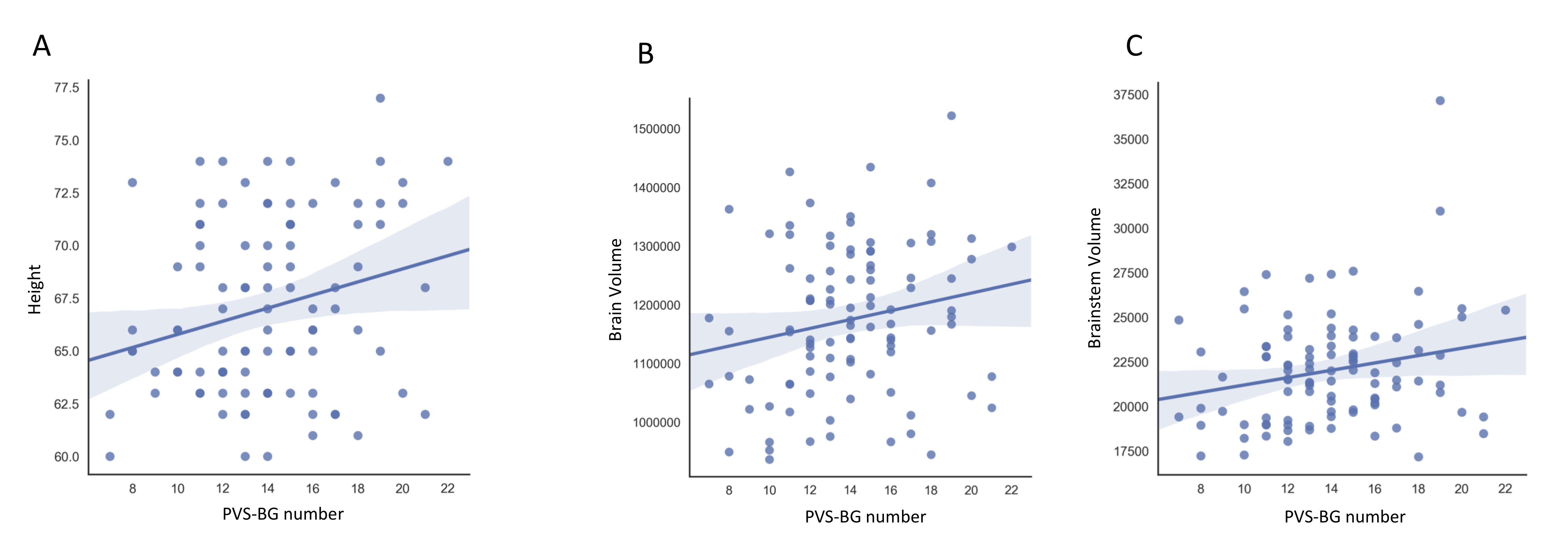
Enlargement of perivascular spaces has been associated with a number of diseases. However, morphological features of perivascular spaces in healthy subjects and their clinical role are still not completely understood. We analyzed on MRI perivascular spaces in a large sample of healthy young subjects. Our results showed a high inter-subjects variability of perivascular spaces. Twins presented similar amount of perivascular spaces. Perivascular spaces in basal ganglia were significantly correlated with subjects’ height, brain volume, and brainstem volume. These findings are relevant for all future studies investigating the role of perivascular spaces.
Introduction
Perivascular spaces (PVS), also known as Virchow-Robin spaces, are tubular fluid-filled structures that surround small blood vessels penetrating the brain parenchyma. In the past, PVS were regarded as having no clinical consequence, but recently they have gained increasing attention from the scientific community thanks to the discovery of the glymphatic system1. Moreover, the improved spatial resolution offered by high- and ultra-high-field MRI is leading to accurate mapping of PVS2. Enlargement of PVS has been associated not only with aging, but also with a number of pathophysiological conditions3. However, physiological imaging characteristics of PVS in healthy subjects are still not clear.Methods
In this study, we analyzed PVS in a large sample of
normal subjects from the Human Connectome Project4. Demographic data and
volume of many brain regions extracted with Freesurfer were collected as well. PVS were independently rated by two
readers on axial T2-weighted images at 3T MRI using a validated 4-point visual
rating scale3. In basal ganglia (PVSBG) and centrum semi-ovale (PVSCS):
0=no PVS, 1=1-10 PVS, 2=11–20 PVS, 3=21–40 PVS, and 4=>40 PVS; in the
midbrain (PVSMB): 0=no PVS, 1=PVS visible. The total PVS score (PVStot)
for each subject was calculated as the sum of the single scores. Results
One hundred healthy subjects (53 females, 47 males; mean age 28.3, range 22-36) were randomly selected from the HCP database. Nine pairs of monozygotic twins were also included in the analysis. The mean number of enlarged PVSCS and PVSBG were 34.7 (range 11-78) and 9.2 (range 3-20), respectively. Each twin showed a similar amount and distribution of PVS compared with the sibling, both in basal ganglia and centrum semiovale. The highest grade of PVSCS was found in 35% of cases; 58% of subjects had enlarged PVS in the midbrain. PVSCS and PVSBG were significantly correlated (p<0.001). Moreover, PVSBG was significantly correlated with subjects’ height (p=0.01), brain volume (p=0.005), and brainstem volume (p=0.005).Discussion & Conclusion
Identifying the physiological factors affecting PVS
in normal subjects is fundamental in order to better understand the
significance of PVS in pathological states and, ultimately, PVS function. This
study demonstrates that a high inter-subjects variability of enlarged PVS is detectable
in a healthy young population. This level of variability was maintained between
the pairs of twins, but the
number of PVS in a twin was comparable to that of the sibling. This finding
suggests a potential genetic contribution to the number and distribution of
PVS. Additionally, more than 1/3 of subjects presented with what is considered
as abnormal level of PVS, highlighting the need for the development of a new
rating scale. We also found that some morphological factors may affect PVS in normal subjects, particularly
in basal ganglia, suggesting the importance of statistical
adjustment for brain size when studying PVS. These results are relevant for
all future studies investigating the role of PVS in pathological conditions and
across different groups.Acknowledgements
No acknowledgement found.References
1. Iliff JJ, Wang M, Liao Y, et al. A paravascular pathway facilitates CSF flow through the brain parenchyma and the clearance of interstitial solutes, including amyloid β. Sci Transl Med. 2012;4(147):1-11. doi:10.1126/scitranslmed.3003748
2. Barisano G, Sepehrband F, Ma S, et al. Clinical 7T MRI: are we there yet? A review about magnetic resonance imaging at ultra-high field. Br J Radiol. October 2018. doi:10.1259/bjr.20180492
3. Potter GM, Chappell FM, Morris Z, Wardlaw JM. Cerebral perivascular spaces visible on magnetic resonance imaging: development of a qualitative rating scale and its observer reliability. Cerebrovasc Dis. 2015;39(3-4):224-231. doi:10.1159/000375153
4. Van Essen DC, Ugurbil K, Auerbach E, et al. The Human Connectome Project: A data acquisition perspective. Neuroimage. 2012;62(4):2222-2231. doi:10.1016/j.neuroimage.2012.02.018
Figures