2558
Assessment of Cerebral Hemodynamics Change of Hypertension using Multi-TI Arterial Spin-LabelingQiuju Fan1, Zhen Yang1, Nan Yu1, Qi Yang1, Yong Yu1, Yue Li1, and Shaoyu Wang2
1Shaanxi University of Chinese Medicine, Xianyang, China, 2MR senior scientific marketing specialist, Siemens Healthineers, Xianyang, China
Synopsis
In our study, we evaluate the diagnostic value of mTI-ASL as a noninvasive method to detect subtle hemodynamic abnormalities in hypertension at different stage.
Purpose
Brain is an important target organ for the harm of hypertension which is associated with microvascular brain injury and induces cerebrovascular remodeling,but its direct influence on the cerebral circulation is not fully clear[1].Our objective was to investigate the hemodynamics changes of whole-brain of different levels of hypertension, using mTI-ASL technique.
Methods
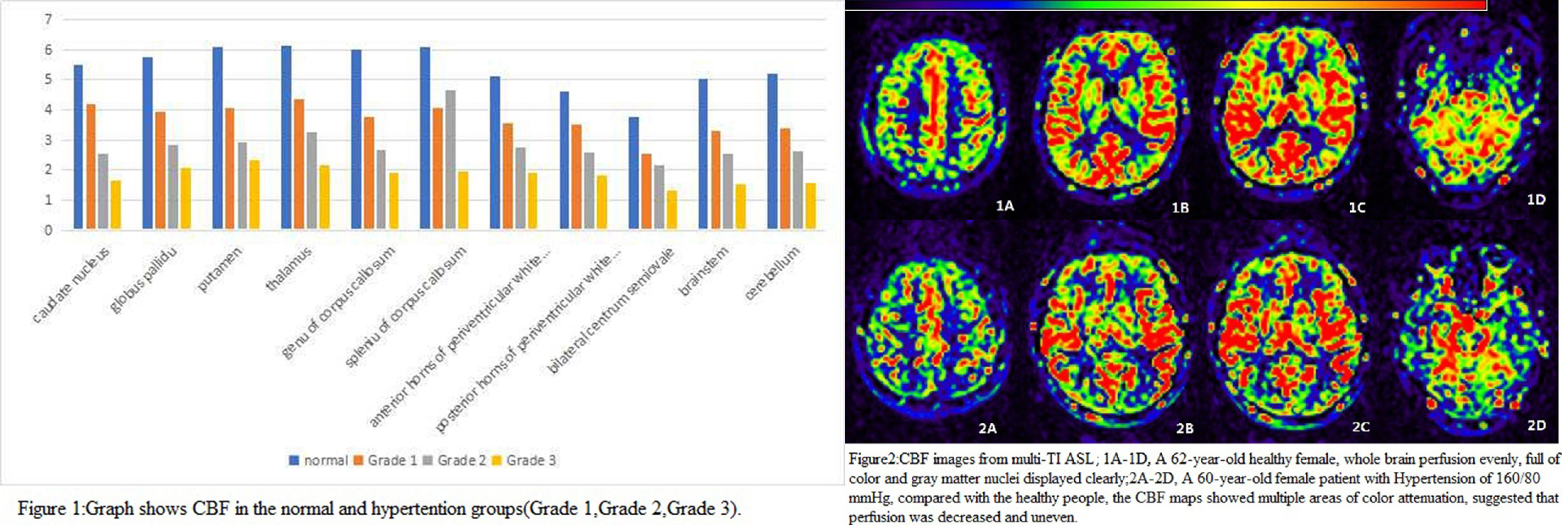
Totally 50 subjects, including a patient group (n=35; 22 males; age= 46.3±7.6 years;test-time blood pressure (BP) =159±25 /99±9mmHg) and an age-matched control group (n=15; 9 males;age=45±6.9 years; test-time BP = 112±11/74±8mmHg) were enrolled in this study and scanned on a 3.0T MRI system (MAGNETOM Skyra, Siemens Healthcare, Erlangen, Germany) using routine MRI and mTI-ASL sequence. The mTI-ASL sequence was used with following parameters: FOV = 220×220 mm2, matrix = 128×128, slice thickness = 4 mm, slices = 30, TR/TE =3200/26 ms, 4 TIs from 500 to 2800 ms,total acquire time 5 min. The patients were divided into three groups according to the systolic blood pressure (SBP);grade 1:140≤SBP≤159mmHg; grade 2: 160≤SBP≤179mmHg; grade 3:SBP≥180mmHg. The cerebral blood flow (CBF) values in various regions of interest (ROIs) were extracted from white matter(bilateral centrum semiovale, anterior and posterior horns of periventricular white matter, genu and spleniu of corpus callosum),caudate nucleus, globus pallidu,putamen, thalamus,brainstem and cerebellum.One-way ANOVA were performed to evaluated the significance of the inte-group difference in CBF modifications.Results
Compared with controls, hypertensive patients exhibited decreased CBF values with the growth of grade of hypertension.The CBF values in varios regions in grade 1 were significantly lower than those in control group except posterior horns of preventricular white matter (P﹥0.05). Furthermore, compared with the grade1 hypertensive patients,grade 2 hypertension showed significantly reduced CBF only in caudate nucleus (P < 0.05).However, it was observed that, between grade 2 and grade 3 hypertensive patients, there were no statistically significant difference in all CBF values (P ﹥0.05).Conclusion
mTI-ASL has the ability to detect subtle hemodynamic abnormalities in hypertension at different stage. Quantitative CBF values usage as a biomarker for disease progression in hypertension is potentially a powerful tool for longitudinal patient monitoring.Acknowledgements
None.References
[1] ME Johnston,K Lu,JA Maldjian,et al. IEEE Transactions on Medical Imaging,34 (2015 ) :1392-1402.Figures
figure