2555
Assessment of Hemodynamic and Hydrodynamic alternations in Spontaneous Intracranial Hypotension by using MR-based Intracranial Pressure Method1Department of Radiology, Taichung Veterans General Hospital, Taichung, Taiwan, 2Department of Education, Taichung Veterans General Hospital, Taichung, Taiwan, 3Department of Medical Imaging and Radiological Sciences, Central Taiwan University of Sciences and Technology, Taichung City, Taiwan, 4Department of Biomedical Engineering, Hung Kuang University, Taichung City, Taiwan
Synopsis
Epidural venous dilatation is commonly seen in patients with spontaneous intracranial hypotension, and its presence often indicates a distinctly altered cerebrospinal hemodynamic/CSF dynamic. With the MR-ICP technique, significant statistical differences were found in various hemodynamic and CSF dynamic parameters. The result suggests that EVD is a representative feature of hemodynamic/CSF-dynamic change in SIH, and also highlights the potential of MR-ICP as a reliable method of assessment for SIH.
Introduction
Spontaneous intracranial hypotension (SIH) presents itself with a great spectrum of symptoms and signs [1]. Amongst them, our previous study presented that the imaging finding of epidural venous dilatation (EVD) in the cervical spine appears to define a subtype of SIH with distinct changes in cerebrospinal hemodynamics [2]. In this study, we statistically analyzed the cerebrospinal hemo- and hydro-dynamics using the MR-intracranial pressure (MR-ICP) scanning technique, to assess the pathophysiological alternations in patients with SIH.Materials and Methods
We recruited 35 patients of SIH, 18 subjects with EVD, 17 subjects without EVD, and 20 normal volunteers were also collected for control. All subjects were scanned with the MR-ICP sequence: phase-contrast MRI (PC-MRI) scans that measures the transcranial arterial and venous blood flow, and oscillatory spinal cerebrospinal fluid (CSF) flow. The details of the MR-ICP method have been previously described by Alperin et al [3]. The peak-to-peak intracranial volumechange (ICVCpp) and CSF pressure gradient (PGcsf-pp) are calcu-lated from the 32 cardiac frames of cerebral blood and CSF pulsa-tile flows. The MR-derived intracranial elastance (IE) index was estimated by dividing PGcsf-pp with ICVCpp, which is based on a monoexponential relationship of the absolute ICP to the derivative of the pressure concerning the volume. The mean MR-ICP indexes and flowparameters were calcu-lated from three consecutive imaging protocols. The statistical significance of differences between groups was calculated using analysis of variance (ANONV) test, with a two-tailed P<0.05.Results
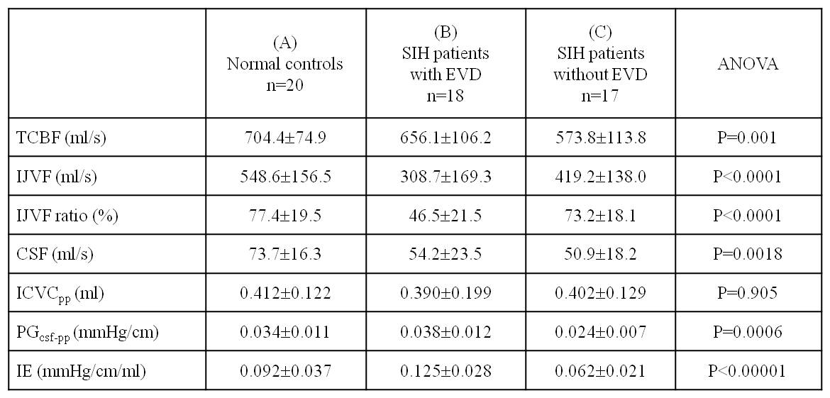
Table 1 shows the ANOVA results of flow parameters and MR-ICP indexes between the two SIH groups and normal controls. It is worth noting that the mean IE, PGcsf-pp, IJVF and IJVF ratios were significantly different in those three groups, with all p-values < 0.0001). The SIH patients with EVD had highest IE and PGcsf-pp, lowest IJVF and IJVF ratio. Contrarily, the SIH patients without EVD had lowest IE and PGcsf-pp. The result would illustrate the shift of venous outflow into the epidural veins would be closely correlated with the changes of the cranio-spinal hydrodynamics and elastance.Discussion
By definition, SIH is a disorder of CSF homeostasis typically resulting from CSF leakage, and therefore low CSF pressure would be presented. However, normal or higher CSF pressure is not uncommon in SIH patients, reported in 13–61% [4]. Although no reference standard was available for validation in the in vivo experiments, our study demonstrated the explicit differences of hemo- and hydro-dynamics in two groups of SIH patients. The decrease in IJVF and IJVF ratio, indicating a more deep-vein shifted hemodynamics, suggest that EVD is likely a presentation of relatively advanced SIH. In addition, the significant difference in IE implies significant alterations in cerebral hemodynamics and CSF dynamics during the physiological process that leads to EVD. These seemingly-contradictory results support the hypothesis that EVD occurs as a compensatory mechanism to maintain equilibrium after the initial change of cerebrospinal hemodynamics and CSF dynamics.Conclusion
Significant statistical differences were found in multiple hemodynamic parameters and MR-ICP indexes among SIH patients with EVD, SIH patients without EVD, and normal volunteers. This result highlights the presence of EVD as a key feature of physiological change in SIH. It also reinforces the reliability of this technique as an assessment method of SIH, and the possibility to further understand this condition in a hemodynamic/CSF-dynamic basis.Acknowledgements
Nil.References
1. Schievink WI. Spontaneous spinal cerebrospinal fluid leaks and intra-cranial hypotension. JAMA 2006;295:2286–2296.
2. Tsai,YH, Chen HC, Tung H, Wu YY, Chen HM, Pan KJ, et al. Noninvasive Assessment of Intracranial Elastance and Pressure in Spontaneous Intracranial Hypotension by MRI. JMRI 2018;48:1255-1263.
3. Alperin N, Lee SH, Loth F, Raksin PB, Lichtor T. MR-intracranial pres-sure (ICP): a method for noninvasive measurement of intracranial pres-sure and elastance: baboon and human study. Radiology 2000;217:877–885.
4. Kranz PG, Tanpitukpongse TP, Choudhury KR, Tanpitukpongse TP,Gray L. How common is normal cerebrospinal fluid pressure in spon-taneous intracranial hypotension? Cephalalgia 2016;36:1209–1217
Figures