2552
Evaluation of Intracranial Pressure-Regulation by MRI-measured Cerebrospinal Fluid PulsationMasatomo Uehara1, Tosiaki Miyati1, Naoki Ohno1, Riho Okamoto1, Moemi Tachimoto1, Mitsuhito Mase2, Hiroshi Furusho1, Satoshi Kobayashi1, and Toshifumi Gabata1
1Division of Health Sciences, Graduate School of Medical Sciences, Kanazawa University, Kanazawa, Japan, 2Department of Neurosurgery, Graduate School of Medical Sciences, Nagoya City University, Nagoya, Japan
Synopsis
We conducted this study to determine the cerebrospinal fluid pulsation in the supine and sitting positions using multiposture MRI. The stroke volume of the aqueduct is not affected by intracranial pressure change. Cerebrospinal fluid pulsation measurements to evaluate the intracranial pressure-regulation function should be taken at the boundary between cranial and spinal cavities rather than in the aqueduct.
INTRODUCTION
Cerebrospinal fluid (CSF) dynamics determine homeostasis and its failure. Numerous studies with phase-contrast magnetic resonance imaging (MRI) have shown CSF pulsation in the aqueduct or in the boundary between cranial and spinal cavities in patients with abnormal CSF dynamics, such as those with idiopathic normal pressure hydrocephalus or with Arnold-Chiari malformation.1-3 However, whether intracranial pressure regulation and compliance can be evaluated by the CSF pulsation measurement alone (particularly in the aqueduct) is unclear. Therefore, we conducted this study to determine the CSF pulsation under different intracranial pressure4,5 (with individuals in the supine and sitting positions) using an original MRI system that can obtain images in any posture (multiposture MRI).6MATERIALS AND METHODS
We obtained velocity-mapped images at the mid-aqueduct level and at the boundary between cranial and spinal cavities (mid-C2 level) in the supine and sitting positions from seven healthy volunteers (aged 22.9 ± 2.0 years) using an ECG-triggered 2D phase-contrast technique with a 0.4-T multiposture MRI machine (HITACHI Healthcare) (Fig. 1a and b). Imaging parameters of the mid-aqueduct scan were as follows: TR, 25 ms; TE, 12.2 ms; flip angle, 8°; R-R interval, 1; velocity-encoded gradient, 7-12 cm/s; imaging matrix, 256 x 128; field of view, 150 x 150 mm. The imaging parameters of the mid-C2 scan were as follows: TR, 25 ms; TE, 9 ms; flip angle, 13°; R-R interval, 1; velocity-encoded gradient, 7-10 cm/s; imaging matrix, 256 x 128; field of view, 160 x 160 mm. Then, we calculated the CSF stroke volume from each scan data and compared those between supine and sitting positions (Fig. 1c and d). Our institutional review board approved the study.RESULTS AND DISCUSSION
The C2 CSF stroke volume in the sitting position (0.67 ± 0.23 mL/min) was significantly lower than that in supine position (1.18 ± 0.22 mL/min, P < 0.05), with a difference of 43.3% ± 17.6% (Fig. 2a), suggesting a decreased need for intracranial pressure-regulation function in the sitting position because of the lower intracranial pressure in this position.4,5 However, we found no significant difference in CSF stroke volumes of the aqueduct between the supine and sitting positions (Fig. 2b), and no significant correlations in the alteration rates of the CSF stroke volume from the supine to the sitting positions between the aqueduct and C2 locations (Fig. 3). Our results indicate that the intracranial cavity pressure-regulation function itself is considerably smaller in magnitude than that of the spinal cavity because of the presence of epidural fat in the spinal canal.CONCLUSION
The stroke volume of the aqueduct is not affected by intracranial pressure change. CSF pulsation measurements to evaluate the intracranial pressure-regulation function should be taken at the boundary between cranial and spinal cavities rather than in the aqueduct.Acknowledgements
No acknowledgement found.References
- Miyati T, Mase M, Tatsuo B, et al. Frequency analysis of CSF flow on cine MRI in normal pressure hydrocephalus. Eur Radiol. 2003; 13: 1019-1024.
- Alperin N, Sivaramakrishnan A, Lichtor T. Magnetic resonance imaging-based measurements of cerebrospinal fluid and blood flow as indicators of intracranial compliance in patients with Chiari malformation. J Neurosurg. 2005; 103: 46-52.
- Miyati T, Mase M, Kasai H, et al. Noninvasive MRI assessment of intracranial compliance in idiopathic normal pressure hydrocephalus. J Magn Reson Imaging. 2007; 26: 274-278.
- Alperin N, Lee SH, Sivaramakrishnan A, et al. Quantifying the effect of posture on intracranial physiology in humans by MRI flow studies. J Magn Reson Imaging. 2005; 22: 591-596.
- Klarica M, Radoš M, Erceg G, et al. The influence of body position on cerebrospinal fluid pressure gradient and movement in cats with normal and impaired craniospinal communication. PLoS One. 2014; 9: e95229.
- Naoki O, Miyati T, Yuki H, et al. Quantitation of venous blood flow in gravity MRI: a phantom study. Medical Imaging and Information Sciences. 2017; 34:141-143.Naoki O, Miyati T, Yuki H, et al. Quantitation of venous blood flow in gravity MRI: a phantom study. Medical Imaging and Information Sciences. 2017; 34:141-143.
Figures
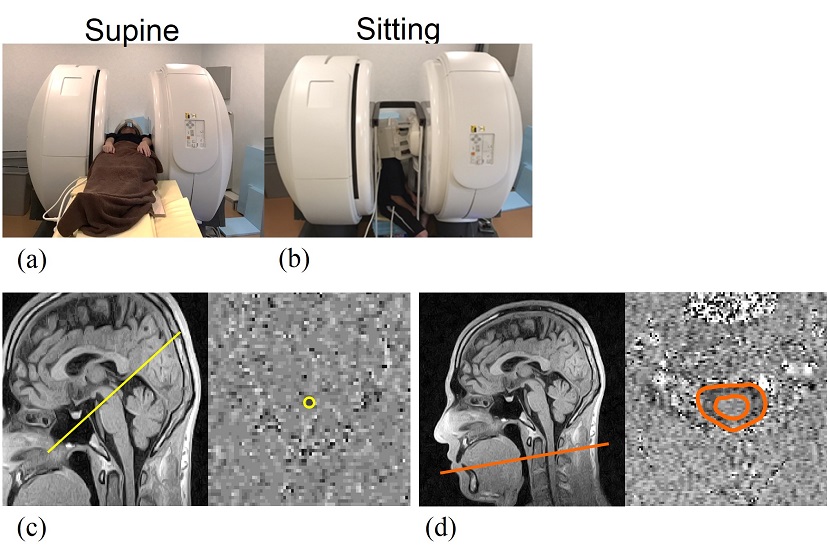
Figure 1. (a) Supine
and (b) sitting positions in the multiposture MRI machine. Imaging planes for mid-aqueduct
scan (left in c) and mid-C2-scan (left in d) on the midsagittal T1-weighted
images, and cerebrospinal fluid (CSF) regions of interest of the aqueduct
(right in c) and the C2 CSF space (right in d) on the phase-contrast velocity
images.
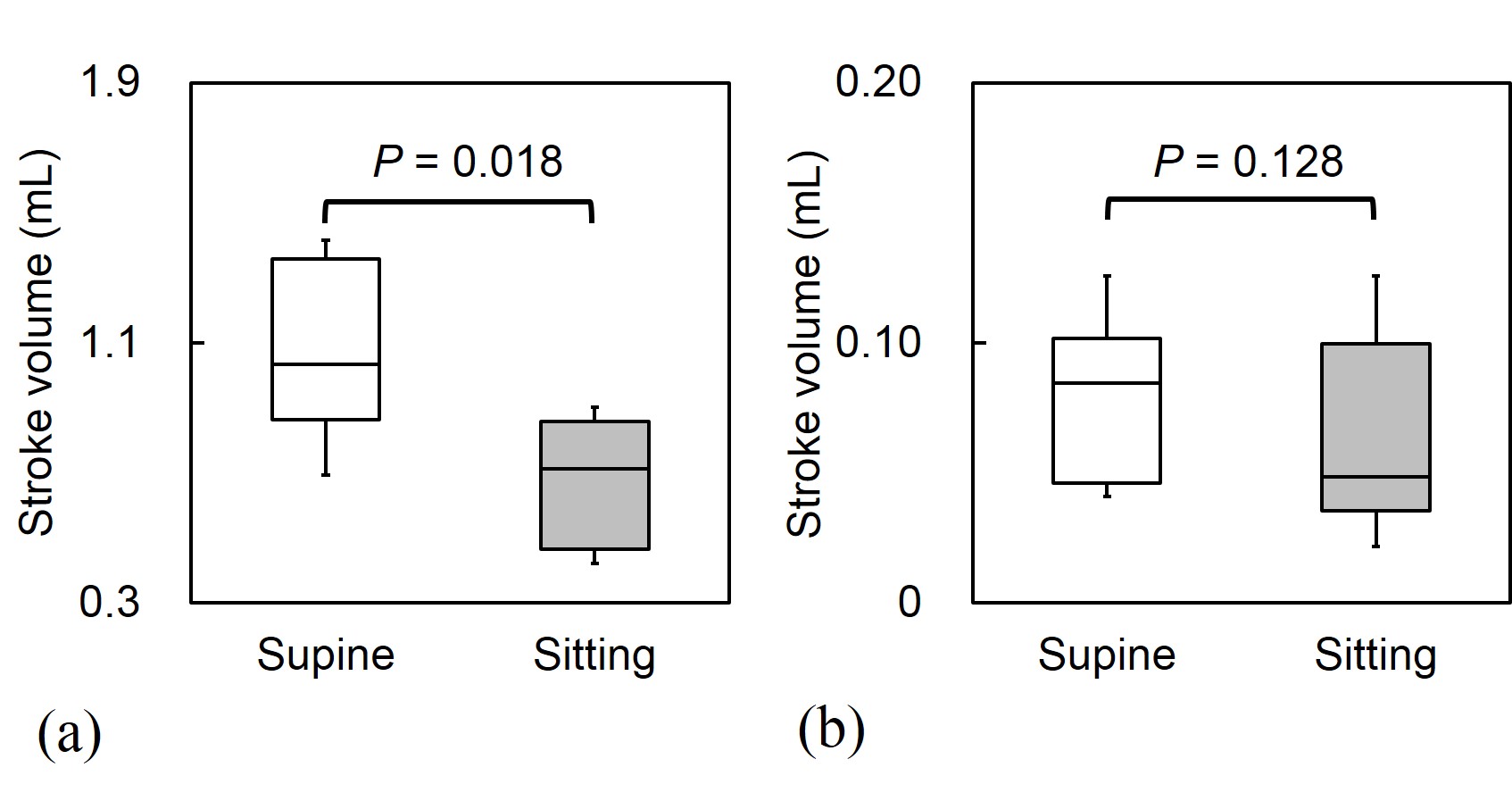
Figure 2. Cerebrospinal
fluid stroke volume of the (a) C2 and (b) aqueduct in the supine and sitting positions.
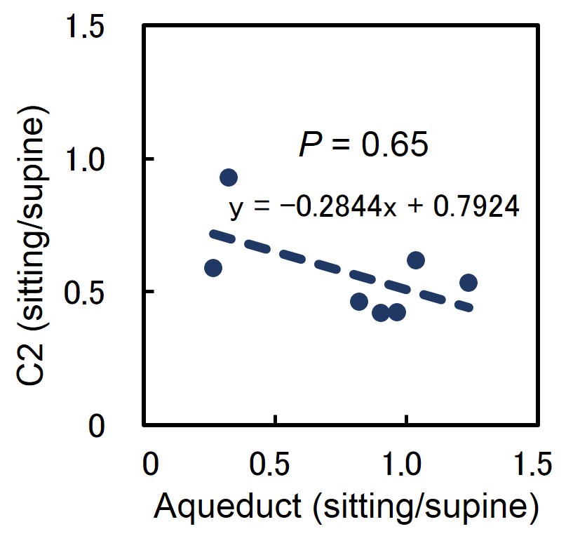
Figure 3. Association
in alteration rates of the CSF stroke volume from the supine to sitting positions between
the aqueduct and C2.